[Preprint]. 2025 Sep 9:rs.3.rs-7512609.
doi: 10.21203/rs.3.rs-7512609/v1.

Oropouche virus disrupts neurodevelopment and is vertically transmitted

Affiliations

Oropouche virus disrupts neurodevelopment and is vertically transmitted

Kellie Jurado et al. Res Sq. .

Abstract

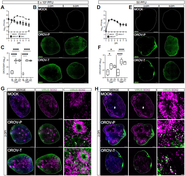
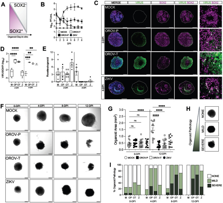
Oropouche virus (OROV) historically caused a self-limiting disease, yet recent strains are associated with congenital infection and neurodevelopmental disruption. These cases highlight a need to study OROV as a congenital pathogen and determine the impact of infection on neurodevelopment. Here, we show that OROV is vertically transmitted and induces a microcephaly-like phenotype in human forebrain organoids. We found OROV robustly infects human neural progenitor cells in organoids. In contrast to ZIKV, OROV had a heightened capacity for infection and organoid pathology. We show this increased pathogenesis is partially attributable to OROV antagonism of innate immune signaling. We further demonstrate that OROV is vertically transmitted and infects the fetal tissues in a murine model of congenital infection. Our results demonstrate that OROV can be vertically transmitted and has heightened capacity for neurodevelopmental disruption. These findings underscore need for monitoring OROV as a re-emerging virus capable of inducing microcephaly in infected fetuses.


Conflict of interest statement

COMPETING INTEREST DECLARATION The authors declare no competing interests.

Figures

Figure 1
Figure 1. OROV infects SOX2+ neural progenitor cells in human forebrain organoids.
Day-in-vitro (DIV) 35 iPSC-derived human forebrain organoids were mock-infected (MOCK or M) or infected with Zika virus (ZIKV or Z), the historical Oropouche prototypical strain (OROV-P or OP), or a recent traveler isolate of Oropouche (OROV-T or OT) at (A-C) 5×10⁴ or (D-F) 50 plaque forming units (PFU) for 24 hours. (A, D) Plaque assays were performed on culture supernatant through 8-days post infection (dpi) to quantify infectious virus production over time. Data presented as mean ± SEM (n ≥ 4 organoids, N ≥ 3 independent experiments). (B, E) Representative immunofluorescent images of organoids mock-infected or infected with OROV-P or OROV-T at 2- or 8-dpi. Organoid sections were outlined using DAPI (blue) and stained for OROV antigen (green). Scale bars = 200 μm. (C, F) Quantification of OROV+ organoid area relative to DAPI+ area in immunofluorescent images. Data presented as box and whisker plot; whiskers indicate minimum and maximum (n ≥ 3 organoids, N ≥ 2 independent experiments). (G-H) Representative immunofluorescent images of mock-, OROV-P- or OROV-T-infected for 2-dpi at (G) 5×10⁴ PFU or (H) 50 PFU. Sectioned organoids were outlined using DAPI (blue) and stained for OROV or ZIKV antigen (green) and SOX2 (magenta). White box indicates inset. Gray arrows indicate virus-infected SOX2+ cells. Scale bars of whole organoids = 200 μm and inset = 20 μm. (n ≥ 4 organoids, N ≥ 2 independent experiments). Statistical analysis performed with one-way ANOVA followed by Sidak’s multiple comparisons test, * p < 0.05, **p < 0.01, ***p < 0.001, ****p < 0.0001.
Figure 2
Figure 2. OROV disrupts forebrain organoid development.
(A) Schematic showing the proportion of SOX2+/SOX2-cells in iPSC-derived human forebrain organoid development. (B-I) DIV15 forebrain organoids were mock-infected or infected with 5×10⁴ PFU of OROV-P, OROV-T, or ZIKV for 24 hours. (B) Viral plaque assay was performed on culture supernatant through 8-days post infection (dpi). Data presented as mean ± SEM. (n ≥ 2 organoids, N ≥ 2 independent experiments). (C) Representative immunofluorescent images of organoids mock-infected or infected with OROV-P, OROV-T at 2-dpi, or ZIKV at 4-dpi. Organoid sections were outlined using DAPI (blue) and stained for OROV or ZIKV antigen (green) and SOX2 neural progenitor cell marker (magenta). White box indicates inset. Gray arrows indicate virus-infected SOX2+ neural progenitor cells. Scale bars of whole organoids = 200 μm and inset = 20 μm. (D) Quantification of OROV+ organoid area relative to DAPI+ area in immunofluorescent images of organoids infected for 2- and 4-dpi. Data presented as box and whisker plot; whiskers indicate minimum and maximum (n ≥ 2 organoids, N ≥ 2 independent experiments). (E) Number of neural rosettes per mock- or virus-infected human forebrain organoid were quantified at 2- and 4-dpi. Data presented as means ± SEM (n ≥ 2 organoids, N ≥ 2 independent experiments). (F) Representative confocal images of mock or virus-infected organoids at 0-, 4-, 8-, and 12-dpi (n ≥ 13 organoids, N = 3 independent experiments). (G) Quantification of mock- or virus-infected organoid area at 0- and 12-dpi. (H) Organoid pathology was assessed via confocal microscopy and categorized as no, mild, or severe pathology. (I) Organoid pathology assessment was quantified and percentages plotted as contingency plots. Differences in categorical distribution of organoid pathology were analyzed using the Cochran-Armitage test and P values were adjusted for multiple comparisons using the Bonferroni correction (n ≥ 13 organoids, N = 3 independent experiments). All other statistical analysis were performed with one-way ANOVA followed by Sidak’s multiple comparisons test, ns p ≥ 0.05, * p < 0.05, ***p < 0.001, ****p < 0.0001.
Figure 3
Figure 3. OROV antagonizes type I interferon signaling.
(A-C) DIV35 forebrain organoids were pretreated with IFNβ or vehicle prior to infection with (A) 5×10⁴ PFU of ZIKV (B) 5×10⁴ PFU or (C) 50 PFU of either OROV-P or OROV-T for 24 hours. Plaque assays were performed on culture supernatant through 8-dpi. Additional IFNβ or vehicle treatments were given at 2-, 4-, and 6-dpi. Data presented as mean ± SEM. (n ≥ 2 organoids, N ≥ 2 independent experiments). (D-E) DIV35 organoids were mock-infected (M) or infected with 5×10⁴ PFU of OROV-P (OP), OROV-T (OT), or ZIKV (Z). Organoid RNA was harvested at 2- and 8-dpi and RT-qPCR was performed for (D) IFNβ and (E) IFIT1. (F) Representative immunofluorescent images of DIV35 forebrain organoids mock-infected, or infected with 5×10⁴ PFU of OROV-P, OROV-T or ZIKV for 24-hours prior to stimulation with IFNβ or vehicle for 24-hours. Organoid sections were outlined using DAPI (blue) and stained for IFIT1 protein (gray), SOX2 neural progenitor cell marker (magenta), and OROV or ZIKV antigen (green). White box indicates inset. Orange arrows indicate virus-infected IFIT1+ cells. Yellow arrows indicate virus-infected IFIT- cells. Gray arrows indicate IFIT+ uninfected cells. Scale bars of whole organoids = 200 μm and inset = 20 μm. (G) Quantification of IFIT1+ area relative to DAPI+ area in immunofluorescent images of organoids in (F). Data presented as box and whisker plot; whiskers indicate minimum and maximum. (n ≥ 7 organoids, N = 2 independent experiments). (H) Quantification of IFIT1+ area that was either OROV negative or OROV positive of organoids in (F). Statistical analysis for (A-C) were performed with ordinary two-way ANOVA followed by Sidak’s multiple comparisons test. All other analyses were performed with one-way ANOVA followed by Sidak’s multiple comparisons test, ns p > 0.05, * p < 0.05, **p < 0.01, ****p < 0.0001.
Figure 4
Figure 4. OROV is vertically transmitted to the developing fetus.
(A) Schematic of experimental design. 9–12-week-old female pregnant C57BL/6 mice (dams) were treated with either 0.5mg/mouse of αIFNAR1 (MAR1–5A3) or isotype control (mouse IgG1) intraperitoneally on embryonic day 5.5 (E5.5). On E6.5, dams were infected with 104 PFU of OROV-P (strain TR9760) subcutaneously via footpad injection. (B-G) All maternal and fetal tissues were analyzed at 7dpi (E13.5). (B) Peripheral maternal tissue (spleen, liver, heart, serum) along with female reproductive tissue (vagina, uterus) were harvested and OROV RNA in peripheral and reproductive maternal tissues quantified via RT-qPCR (n=3 dams per condition, N=3 independent experiments). (C) Representative images of matched fetuses and placentas from isotype-and αIFNAR1-treated dams. (D) Quantification of resorbed and intact fetal tissue (E) Quantification of fetal body area. (F) OROV RNA in placenta and fetal heads were quantified via RT-qPCR. Open symbols denote fetal resorptions. (G) Viral titers in placenta and fetal heads were quantified via plaque assay. Dashed line indicates limit of detection for each assay (n>14 samples per condition, N=3 independent experiments). Statistical analysis for (F) was performed with an unpaired t-test. All other analyses were performed with two-way ANOVA adjusted for multiple comparisons, ns p > 0.05, * p < 0.05, ***p < 0.001, ****p < 0.0001.

References

    1. Sakkas H., Bozidis P., Franks A. & Papadopoulou C. Oropouche Fever: A Review. Viruses 10, 175 (2018). - PMC - PubMed
    1. Elliott R. M. Orthobunyaviruses: recent genetic and structural insights. Nat. Rev. Microbiol. 12, 673–685 (2014). - PubMed
    1. Medeiros de D. B.A. et al. Case Series of Adverse Pregnancy Outcomes Associated with Oropouche Virus Infection. Viruses 17, 816 (2025). - PMC - PubMed
    1. Gatti F. D. et al. Neuroinvasive Oropouche virus in a patient with HIV from extra-Amazonian Brazil. Lancet Infect. Dis. (2025) doi: 10.1016/s1473-3099(25)00352-4. - DOI
    1. Ewing C. B., Siraj D. & Lepak A. First Case of Neuroinvasive Oropouche Virus in Wisconsin: A Case Report. Open Forum Infect. Dis. ofaf402 (2025) doi: 10.1093/ofid/ofaf402. - DOI - PMC - PubMed

Publication types

LinkOut - more resources