[Preprint]. 2025 Sep 8:2025.09.07.674757.
doi: 10.1101/2025.09.07.674757.

Febrile temperature enhances Plasmodium falciparum cytoadhesion by disrupting the endothelial glycocalyx

Affiliations

Febrile temperature enhances Plasmodium falciparum cytoadhesion by disrupting the endothelial glycocalyx

Viola Introini et al. bioRxiv. .

Abstract

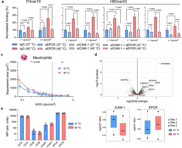
Fever, a universal host defense in infection and inflammation, paradoxically contributes to neurological complications in malaria. While febrile temperatures enhance the expression of parasite virulence proteins that mediate vascular adhesion and disease severity, its effects in the endothelium remain elusive. Here we present a 3D fever-on-a-chip model that recapitulates human brain and lung microvessels under febrile conditions. Short febrile episodes at 40 °C, common in treated cerebral malaria patients, rapidly enhanced iRBC and immune cell binding under flow. Mechanistically, we demonstrated that this phenotype was driven by endothelial glycocalyx shedding, which exposed endothelial receptors EPCR and ICAM-1. Preserving glycocalyx integrity with a broad MMP inhibitor prevented the temperature-induced rise in cytoadhesion. These findings identify fever as a host-specific amplifier of vascular pathology in malaria and highlight endothelial-protective or antipyretic interventions as important strategies to mitigate febrile microvascular pathology.


Conflict of interest statement

Competing interests The authors declare no competing interests.

Figures

Fig. 1.
Fig. 1.. Fever quantification in paediatric malaria patients and P. falciparum-iRBC binding at 37 and 40 °C to 3D microvessels.
a, Distribution of maximum body temperature data during admission from 146 paediatric malaria cases in Zambia and Malawi treated with antipyretics as described in Methods. b, Duration of high fever episodes ≥ 40 °C or between 39 and 40 °C. Bar plot represent median and interquartile range; μ indicates group mean. c, Simulated mid-plane flow velocity in the grid network prior to collagen remodelling by HBMECs (see Methods). Inset: lumen cross-section velocity and WSS at first branch (black line).d, Volumetric reconstruction of a microvessel network grid cross section labelled with VE-cadherin antibody (magenta) and nuclear staining by DAPI (blue). Scale bar = 50 μm. e, Schematic representation of the binding assay workflow. f-i, Receptor binding schematic and iRBC sequestered areas at 37 °C and 40 °C across WSS for: f, HB3var03; g, IT4var19; h, IT4var31; i, NF54var2csa. Medians are represented by dots and interquartile range by error bars. Statistical analysis of binned regions (< 1 dyn/cm2 and ≥ 1 dyn/cm2) (dotted line) was determined by Mann-Whitney U test (n = 6 independent biological replicates for HB3var03, IT4var19, and IT4var31; n = 4 for NF54var2csa). Scale bars = 200 μm.
Fig. 2.
Fig. 2.. Receptor-mediated binding of P. falciparum and neutrophils at febrile temperature occurs without altering endothelial protein level.
a, Binding of IT4var19 strain iRBCs at 37 °C or 40 °C in the presence of IgG isotype control (filled) or anti-EPCR mAb 252 antibody (stripes). b, Binding of HB3var03 strain iRBCs at 37 °C or 40 °C in the presence of IgG isotype control (filled) or both anti-EPCR mAb 252 (stripes), anti-ICAM-1 mAb 15.2 (squares), and both (light colour). Percentage of binding for each treatment is normalised to the respective IgG control at 37 °C. Bars represent mean ± SEM. Statistical analysis for the two WSS regions WSS <1 dyn/cm2 and WSS ≥ 1 dyn/cm2 using Mann-Whitney U test; n=4–6 independent microvessels. Stratified data can be found in Extended Fig. 5. c, Sequestered neutrophil area at 37 °C and 40 °C across WSS (medians as lines and interquartile ranges as error bars). Statistical analysis for the two WSS regions WSS <1 dyn/cm2 and WSS ≥ 1 dyn/cm2 using Mann-Whitney U test; n = 4 biological replicates per condition. d, Volcano plot of differential protein expression of HBMEC monolayer exposed to 37 °C or 40 °C for 1 h (top), with labeled candidates showing statistically significant changes in protein expression. ICAM1 and EPCR differential protein expression (bottom). n = 3 independent biological replicates. e, MFI of HBMEC surface level expression by flow cytometry after 1h at 37 °C and 40 °C. Bars represent mean ± standard deviation (SD) (n = 5–6 biological replicates).
Fig. 3.
Fig. 3.. Febrile temperature induces shedding of the endothelial glycocalyx.
Immunofluorescence z-stack of microvessel cross sections (left) and MFI normalised by number of cells quantified by DAPI (left) of a, sialic acids labelled with FITC-conjugated Wheat Germ Agglutinin (WGA), b, heparan sulfate and c, syndecan-4; box plots show mean ± SD, n = 4 microvessels per condition, each dots is a region of interest. Scale bars: 50 μm. d, Syndecan-4 shedding quantified by ELISA of 3D brain microvessel supernatants following exposure to 37 °C or 40 °C. e, Syndecan-1 shedding quantified by ELISA of 3D pulmonary microvessel supernatants. Graphs show mean ± SD, which each dot is from a different microvessel batch. Immunofluorescence MFI quantification of f, sialic acids labelled with WGA, and g, heparan sulfate in 3D pulmonary microvessels; box plots show mean ± SD, n = 4 microvessels per condition, each dots is a region of interest. Conditions in a-g include 37 °C or 40 °C incubation for 1 hour, and NA treatment for 30 min at 37 °C (1 U/mL). Statistical analysis was done by one-way ANOVA with Tukey’s multiple comparisons or pairwise comparisons were performed using the two-sided Mann-Whitney U test. h-i) HB3var03 and IT4var19 binding to 3D brain microvessels treated with NA (n = 5 microvessels) compared to 37 °C and 40 °C (n = 6 microvessels). Medians are shown as dots; error bars represent interquartile range. Statistical analysis by Kruskal-Wallis test for binned WSS regions (< 1 dyn/cm2 vs ≥ 1 dyn/cm2; dotted line).
Fig. 4.
Fig. 4.. Analysis of MMP1 secretion, P. falciparum-iRBC binding and endothelial glycocalyx protection after treatment with the MMP inhibitor, batimastat
a, Quantification of angiogenesis-related molecule secretion from HBMEC monolayers after 1 h at 37 °C or 40 °C. Supernatants pooled from n=3 biological replicates and normalised as described in Methods. b-c, HB3var03 and IT4var19 binding after exposure of batimastat-pre-treated microvessels to 37 °C or 40 °C (n = 4 brain microvessels). Median ± error bars representing interquartile range. Statistical analysis by Kruskal-Wallis test for binned WSS regions (< 1 dyn/cm2 vs ≥ 1 dyn/cm2; dotted line). d-f, Z-projection immunofluorescence images of batimastat pre-treated microvessel junctions exposed to 37 °C or 40 °C and labelled for WGA, heparan sulfate, syndecan-4 and DAPI (cyab) (left) and measured glycocalyx marker MFI normalised by number of cells using DAPI (right) (n = 4 brain microvessels, dots represent quanfied ROI). Statistical analysis by one-way ANOVA with Tukey’s multiple comparisons; box plots show mean ± SD. Scale bars = 50 μm.

References

    1. Yamagata K. et al. Coexpression of Microsomal-Type Prostaglandin E Synthase with Cyclooxygenase-2 in Brain Endothelial Cells of Rats during Endotoxin-Induced Fever. J Neurosci 21, 2669–2677 (2001). - PMC - PubMed
    1. Eskilsson A. et al. Immune-Induced Fever Is Dependent on Local But Not Generalized Prostaglandin E2 Synthesis in the Brain. J. Neurosci. 37, 5035–5044 (2017). - PMC - PubMed
    1. Lazarus M. et al. EP3 prostaglandin receptors in the median preoptic nucleus are critical for fever responses. Nat Neurosci 10, 1131–1133 (2007). - PubMed
    1. Evans S. S., Repasky E. A. & Fisher D. T. Fever and the thermal regulation of immunity: the immune system feels the heat. Nat Rev Immunol 15, 335–349 (2015). - PMC - PubMed
    1. Launey Y., Nesseler N., Mallédant Y. & Seguin P. Clinical review: fever in septic ICU patients--friend or foe? Crit Care 15, 222 (2011). - PMC - PubMed

Publication types

LinkOut - more resources