

This is a preprint.
It has not yet been peer reviewed by a journal.
The National Library of Medicine is
running a pilot
to include preprints that result from research funded by NIH in PMC and PubMed.
[Preprint]. 2025 Sep 8:2025.09.08.674823.
doi: 10.1101/2025.09.08.674823.
De novo design of semisynthetic protein nanopores
Affiliations
- PMID: 40964308
- PMCID: PMC12439879
- DOI: 10.1101/2025.09.08.674823
Item in Clipboard
De novo design of semisynthetic protein nanopores
bioRxiv.
.
Abstract
Protein nanopores are essential components of single-molecule oligonucleotide sequencing and sensing devices. Here, we demonstrate that installing additional de novo subunits enables large-scale architectural changes of nanopore complexes. We design de novo proteins that integrate seamlessly with the CsgG pore to form 18-subunit, 315-kilodalton complexes with precisely sculpted pore architectures and tailored ion conduction, opening new possibilities for engineering enhanced nanopores with customized structural and functional properties.
Figures
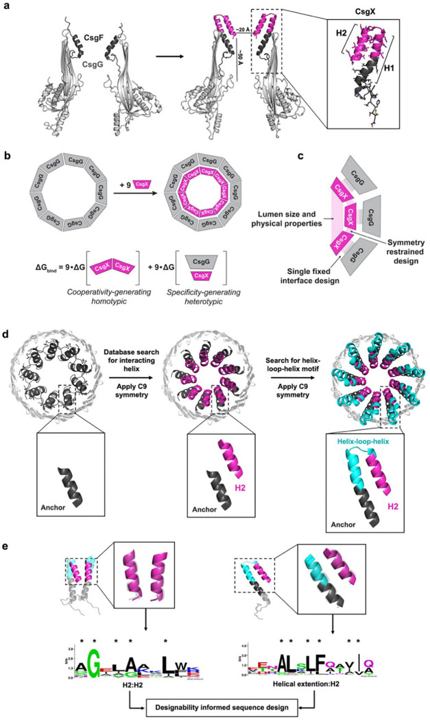
a, Schematic of pore engineering. Starting from the (CsgG:CsgF)9 complex (left), a de novo designed helical hairpin (CsgX, magenta) is precisely grafted onto the C-terminus of CsgF (dark grey), allowing extension into the CsgG lumen (light grey). The design begins with the Anchor helix from CsgF (dark grey) and the de novo portion includes an extension of the Anchor helix to afford H1, a loop, and a second helix H2 (magenta). The boxed inset shows the resulting CsgX monomer (magenta). b, Schematic of pore assembly of CsgX (magenta) with CsgG9 (light grey). The binding energy (ΔGbind) is the sum of homotypic (CsgX:CsgX) and heterotypic (CsgG:CsgX) interactions over all subunits. c, Key structural features that needed to be accommodated during design include pore lumen dimensions, symmetry, and the fixed structure and sequence of (CsgG)9. d, Design approach. Starting from the CsgF Anchor (left), a single helix is geometrically docked against the Anchor and propagated by C9 symmetry (magenta; middle). MASTER is then used to identify geometrically compatible helical extensions of the Anchor and an adjoining helix, creating a helix–loop–helix motif (cyan; right). Candidate motifs are/were? filtered for structural clashes and a cone-like overall structure, funneling to a ~ 20 Å restriction. e, The prevalence of the helical interface geometries across the PDB is evaluated using MASTER to identify frequently occurring, designable motifs. These motifs (transparent grey, overlaid on the H1:H2 and Helical extention:H2 helices) are used to generate sequence logos that capture position-specific residue preferences (right), which are considered as restraints for Rosetta-based sequence design (residue seeds are marked with an asterisk).

a, Blue native gel electrophoresis of the CsgG:CsgX assemblies confirm that all six of the CsgX variants assembled into homogeneous 9:9 complexes with CsgG, with no detectable sub-stoichiometric intermediates. b, e, h, j, Single-channel recordings of (CsgG:CsgX)9 complexes. Representative current traces at 180 mV from synthetic membrane recordings of the (CsgG:CsgX)9 complexes inserted into a MinION flow cell from Oxford Nanopore Technologies. Current/time traces were collected by measuring the current with a frequency of 4kHz. The individual values of the current were analyzed using a normal distribution, giving a mean current and an associated standard deviation. The theoretical curves from fitting the data for(CsgG)9 and (CsgG:CsgF)9 overlayed on each of the (CsgG:CsgX)9 plots in light blue dashed lines (105 pA and 175 pA, respectively) for reference (c,d, CsgG9 and (CsgG:CsgF)9, f,g, (CsgG:CsgX1cyc)9 and (CsgG:CsgX1)9, i,l, (CsgG:CsgX2cyc)9 and (CsgG:CsgX2)9, k,m, (CsgG:CsgX3cyc)9 and (CsgG:CsgX3)9). n, Representative 2D class averages (left) highlighting views of the single nanopore assembly (CsgG:CsgX1)9 and clipped 3D volume of the reconstructed density map for (CsgG:CsgX1)9, low-pass filtered to 15 Å, displayed at contour of 0.06 in UCSF ChimeraX. Density for CsgG and CsgX1 are colored grey and magenta, respectively. o, Low pass filtered 3D volume (transparent grey/magenta) overlaid on the structural model of (CsgG:CsgX1)9 shows unambiguous density for the full CsgX1 hairpin and for all nine CsgX1 monomers in the assembly (end view, left; clipped side view, right). p, Comparison of design model (grey) to structure (magenta) of 3 CsgX1 monomers, with side chains displayed for key polar interactions (R46, D43, D45; top) and hydrophobic packing residues (L48, L50, F51, I52; bottom). q, Alignment of design (gray) to structure (magenta) of designed residues (31–57) of (CsgX1)9 show low overall RMSD (< 1 Å). r, In the full (CsgG:CsgX1)9 structure, the CsgX1 hairpin (magenta) is shifted (~15°) about the axis of symmetry relative to the design model (gray), while retaining the overall pore geometry. s, 3D volumes of (CsgG:CsgX2)9 and (CsgG:CsgX2cyc)9, low-pass filtered to 15 Å, contoured to 0.09 in UCSF ChimeraX. CsgG and CsgX2 shown in transparent grey and purple, respectively, and overlaid on (CsgG:CsgX2)9 design model, show that the (CsgG:CsgX2)9 volume has no observable density for the designed hairpin, while (CsgG:CsgX2cyc)9 has clear density for the full CsgX2. t, Comparison of design model (grey) to structure (purple) of 3 CsgX2cyc monomers, with side chains displayed for key polar interactions (S49, Q52, T53; left) and hydrophobic packing residues (L50, I54, I55; right). u, RMSD of the peptide Cα atoms over a 200 ns molecular dynamics (MD) production run of the designed (CsgG:CsgX)9 models. The initial 20 ns of the 220 ns simulation were excluded from analysis, and RMSD values were calculated relative to the structure at 20 ns. Over the final 50 ns of the simulation, (CsgG:CsgX2)9 (solid purple) exhibited a higher rate of RMSD change (2.82 Å/μs) compared to the other samples (1.69 Å/μs, 2.43 Å/μs, and 1.11 Å/μs for (CsgG:CsgX1)9 (solid pink), (CsgG:CsgX1cyc)9 (dashed pink), and (CsgG:CsgX2cyc)9 (dashed purple), respectively).
References
Publication types
Grants and funding
LinkOut - more resources
Full Text Sources