A nationwide cross-sectional study on endodontic instrument fractures and development of a comprehensive classification system
- PMID: 40964639
- PMCID: PMC12440341
- DOI: 10.4103/JCDE.JCDE_307_25
A nationwide cross-sectional study on endodontic instrument fractures and development of a comprehensive classification system
Abstract
Aim: To assess the incidence and characteristics of instrument fractures in root canals and provide researchers with a systematic classification system that will facilitate diagnosis, research, and treatment planning.
Materials and methods: In this cross-sectional and observational study, we collected the data on 887 intraoral periapical (IOPA) radiographs taken from different geographical and clinical practices in India. The images containing instrument fractures were classified according to the location of fractures within the canal (coronal, middle, and apical third) and their relationship to canal curvature. The IOPA images were independently evaluated by two experienced endodontists, and the kappa statistics was applied to estimate interobserver agreement (≥80%). Descriptive statistics were analyzed using the SPSS software version 22.0.
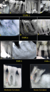
Results: The location of 54.3% of fractures was in the middle third of the canal, followed by apical thirds (24.5%) and coronal thirds (19.8%). Fractures beyond the periapex were rare (1.4%). Of the fractures, 54.3% had curvatures of the canal and of those, 33.1% or 3.1% of the fractures occurred within mild to moderate curvatures. The study found statistically significant differences in fractures in the different regions of the canal (P < 0.01). A unique classification system was created to categorize fractures by location, extent, and curvature involvement which can be used and applied in both clinical and research settings.
Conclusions: The research draws the attention to the lack of a standardized classification system for instrument fractures, an underappreciated aspect of endodontics. The classification system proposed here will enhance communication, educate on diagnosis, treatment planning, and provide support for advanced education in the field.
Keywords: Endodontic instrument fracture; endodontic instrumentation; new classification; root canal treatment.
Copyright: © 2025 Journal of Conservative Dentistry and Endodontics.
Conflict of interest statement
There are no conflicts of interest.
Figures


References
-
- Gulabivala K, Ng YL. Factors that affect the outcomes of root canal treatment and retreatment-a reframing of the principles. Int Endod J. 2023;56(Suppl 2):82–115. - PubMed
-
- Chaniotis A, Ordinola-Zapata R. Present status and future directions: Management of curved and calcified root canals. Int Endod J. 2022;55(Suppl 3):656–84. - PubMed
-
- Ungerechts C, Bårdsen A, Fristad I. Instrument fracture in root canals –Where, why, when and what?A study from a student clinic. Int Endod J. 2014;47:183–90. - PubMed
LinkOut - more resources
Full Text Sources
