Multicenter Study
. 2025 Sep;21(9):e70659.
doi: 10.1002/alz.70659.

Periventricular diffusivity reflects APOE ε4-modulated amyloid accumulation and cognitive impairment in the Alzheimer's disease continuum

Affiliations
Multicenter Study

Periventricular diffusivity reflects APOE ε4-modulated amyloid accumulation and cognitive impairment in the Alzheimer's disease continuum

Chang-Le Chen et al. Alzheimers Dement. 2025 Sep.

Abstract

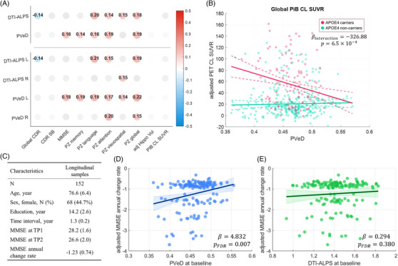
Introduction: Altered glymphatic-related fluid dynamics are increasingly recognized as a feature of Alzheimer's disease (AD). We generalized an established diffusion imaging framework to quantify periventricular diffusivity (PVeD), hypothesizing that fast diffusion signals in the periventricular region can reflect amyloid beta (Aβ) deposition across the AD continuum.

Methods: Participants from two multi-site cohorts (n = 440 and 414), comprising cognitively unimpaired individuals, those with mild cognitive impairment, and patients with AD, were included. We tested and validated the association of PVeD with Aβ burden and core AD characteristics.

Results: Lower PVeD was extensively associated with greater Aβ burden, neurodegeneration, cognitive impairment, and clinical severity in the clinical cohort. Importantly, the relationship between PVeD and Aβ burden was significantly modulated by apolipoprotein E (APOE) ε4 status; APOE ε4 carriers exhibited a replicable stronger negative association. Baseline PVeD also predicted longitudinal cognitive decline.

Discussion: These findings suggest that periventricular diffusion signals reflect APOE ε4-modulated Aβ burden and cognitive decline in AD.

Highlights: An automated method for quantifying periventricular diffusivity (PVeD) is developed. Lower PVeD is associated with higher amyloid load only in a mild cognitive impairment-dominant cohort. Higher amyloid burden may mediate the link between lower PVeD and poorer cognitive outcomes in the clinical cohort. Apolipoprotein E ε4 carriers show a reproducibly stronger inverse PVeD-amyloid association than non-carriers. Baseline PVeD can predict longitudinal Mini-Mental State Examination decline in two independent cohorts.

Keywords: Alzheimer's disease; amyloid imaging; apolipoprotein E ε4; cognitive decline; dementia; diffusion tensor image analysis along the perivascular space; diffusion tensor imaging; diffusivity; perivascular space; periventricular area.


Conflict of interest statement

The authors declare that they have no financial/non‐financial and direct/potential conflicts of interest. Author disclosures are available in the supporting information.

Figures

FIGURE 1
FIGURE 1
Overview of the study. A conceptual framework of fluid dynamics describes how, within the glymphatic system, solutes such as metabolic waste are propelled toward the perivenous conduit, accompanied by CSF and ISF exchange from the brain parenchyma. This study builds on the method proposed by Taoka et al., who introduced DTI‐ALPS to estimate diffusion‐based proxies related to glymphatic integrity given this framework. Rather than targeting predefined regions of interest, we developed an automated approach to delineate the periventricular region using DTI data. We then calculated the TTR on a voxel basis to capture the transverse component of fast diffusion signals within the periventricular region, which primarily contains deep medullary veins oriented along the left‐right axis. We hypothesize that such PVeD can reflect glymphatic‐related activities such as Aβ deposition. To test this, we analyzed two multi‐site, multi‐modal datasets that included CU individuals, individuals with MCI, and patients with AD and other types of dementia for exploration and replication. Our investigation focused on (1) the association of PVeD with Aβ burden, symptom severity, neurodegeneration, and cognitive function, and (2) the modulatory effect of APOE ε4 on the association between PVeD and Aβ burden. Additionally, we tested whether the baseline PVeD metrics can predict longitudinal cognitive change. Aβ, amyloid beta; AD, Alzheimer's disease; APOE, apolipoprotein E; CDR‐SB, Clinical Dementia Rating Sum of Boxes; CL, Centiloid; CSF, cerebrospinal fluid; CU, cognitively unimpaired; DTI, diffusion tensor image; DTI‐ALPS, diffusion tensor image analysis along the perivascular space; ISF, interstitial fluid; MCI, mild cognitive impairment; MD, mean diffusivity; MMSE, Mini‐Mental State Examination; PET, positron emission tomography; PVeD, periventricular diffusivity; SWI, susceptibility‐weighted imaging; T1w, T1‐weighted imaging; TTR, transverse tensor ratio.
FIGURE 2
FIGURE 2
Flowchart illustrating the proposed method for estimating the transverse portion of PVeD. DWIs were preprocessed including phase distortion, eddy current, and motion corrections (A). The corrected DWIs were reconstructed using QSDR to map images into the MNI space and estimate diffusion tensors (B). Meanwhile, MD and axis‐specific diffusivity maps (Dxx, Dyy, and Dzz) were calculated (C). By using axis‐specific diffusivity maps, a novel TTR contrast was defined to reflect transverse water diffusivity potentially along the perivascular space in deep medullary veins (C). PVeA was identified using an automated region growing algorithm applied to MD maps, incorporating lateral ventricle pre‐mask (pre‐LV mask) and anatomically guided dilation (D). The final PVeA mask was intersected with white matter masks, and average TTR values within the PVeA mask were calculated to approximate the apparent PVeD (E), which is used as an overall metric to represent interstitial fluid properties that could be related to glymphatic function. DWI, diffusion‐weighted image; LV, lateral ventricle; MD, mean diffusivity; MNI, Montreal Neurological Institute; PveA, periventricular area; PVeD, periventricular diffusivity; QSDR, q‐space diffeomorphic reconstruction; TTR, transverse tensor ratio.
FIGURE 3
FIGURE 3
Partial correlation matrix of diffusion imaging‐derived metrics with multifaceted clinical characteristics. Diffusion imaging‐derived metrics refer to the DTI‐derived measures including DTI‐ALPS, PVeD, FA, and MD. The multifaceted clinical characteristics include four domains: symptom severity, cognitive outcomes, neurodegeneration, and Aβ burden. The matrix was generated using the BICWALZS dataset (n = 440). Panels (A) and (B) present the correlation matrix of overall diffusion imaging‐derived metrics and that of bilateral metrics for DTI‐ALPS and PVeD, respectively. Values are Spearman correlation coefficients for each pairwise correlation. Covariates including age, sex, and education were adjusted. Hippocampal volume was additionally adjusted by the total intracranial volume. Darker blue and red colors indicate greater negative and positive correlations, respectively. Gray dots indicate those did not pass the significance threshold adjusted by multiple comparisons (adjusted alpha threshold = 0.0031). Aβ, amyloid beta; adj, adjusted; avg, average; BICWALZS, Biobank Innovations for Chronic Cerebrovascular Disease with Alzheimer's Disease Study; CDR, Clinical Dementia Rating; CDR‐SB, Clinical Dementia Rating Sum of Boxes; Cing, cingulum; CL, Centiloid; CU, cognitively unimpaired; DTI, diffusion tensor imaging; DTI‐ALPS, diffusion tensor image analysis along the perivascular space; Front, frontal; FA fractional anisotropy; Hippo, hippocampal; K‐BNT, Korean version of the Boston Naming Test; K‐CWST, Korean version of the Color Word Stroop Test; L, left; MCI, mild cognitive impairment; MD, mean diffusivity; MMSE, Mini‐Mental State Examination; MTA, medial temporal lobe atrophy; Occi, occipital; Parie, parietal; PET, positron emission tomography; PVeD, periventricular diffusivity; R, right; RCFT, Rey Complex Figure Test and Recognition Trial; SUVR, standardized uptake value ratio; SVLT E, Seoul Verbal Learning Test Elderly version; Temp, temporal; Vol, volume; WM, white matter; z, z score.
FIGURE 4
FIGURE 4
Aβ burden mediates the relationship between PVeD and cognitive decline in the BICWALZS cohort. Aβ burden is quantified using the global amyloid PET SUVR, and cognitive decline is represented by MMSE scores and CDR‐SB scores. Panels (A), (B), (C), and (D) illustrate the conditions of the mediation analysis for the following variable pairs: PVeD versus MMSE, PVeD versus CDR‐SB, DTI‐ALPS versus MMSE, and DTI‐ALPS versus CDR‐SB, respectively. Unstandardized coefficients are reported, and the PM represents the indirect effect relative to the total effect. Multiple comparisons are controlled using the Benjamini–Hochberg correction accounting for the number of imaging features tested. Aβ, amyloid beta; BICWALZS, Biobank Innovations for Chronic Cerebrovascular Disease with Alzheimer's Disease Study; CDR‐SB, Clinical Dementia Rating Sum of Boxes; CL, Centiloid; DTI‐ALPS, diffusion tensor image analysis along the perivascular space; FDR, false discovery rate; MMSE, Mini‐Mental State Examination; PET, positron emission tomography; PM, proportion of mediation; PVeD, periventricular diffusivity; SUVR, standardized uptake value ratio.
FIGURE 5
FIGURE 5
The presence of the APOE ε4 allele enhances the association between PVeD and Aβ deposition in the BICWALZS cohort. The amyloid PET CL SUVRs were sampled from five anatomical regions including the cingulum (A), frontal lobe (B), parietal lobe (C), occipital lobe (D), and temporal lobe (E), with the inclusion of the global measure (F). The models included age, sex, and education as covariates. Multiple comparisons were controlled using the Benjamini–Hochberg correction accounting for the number of image measures analyzed. Green and red colors represent APOE ε4 non‐carriers and APOE ε4 carriers, respectively. The interaction terms between PVeD and APOE ε4 are reported. Aβ, amyloid beta; APOE, apolipoprotein E; BICWALZS, Biobank Innovations for Chronic Cerebrovascular Disease with Alzheimer's Disease Study; CL, Centiloid; FDR, false discovery rate; PET, positron emission tomography; PVeD, periventricular diffusivity; SUVR, standardized uptake value ratio.
FIGURE 6
FIGURE 6
Higher baseline PVeD is associated with better longitudinal cognitive outcomes in the BICWALZS cohort. The annual change rate of MMSE is used as the dependent variable with the baseline PVeDs including left PVeD (A), right PVeD (B), and mean PVeD (C) as the primary independent variable. Covariates include baseline age, sex, and education. The Benjamini–Hochberg correction was applied to account for multiple comparisons given the number of imaging measures analyzed. For reference, the analysis was also performed using DTI‐ALPS metrics including left DTI‐ALPS (D), right DTI‐ALPS (E), and mean DTI‐ALPS (F). The coefficients of slope are reported. BICWALZS, Biobank Innovations for Chronic Cerebrovascular Disease with Alzheimer's Disease Study; DTI‐ALPS, diffusion tensor image analysis along the perivascular space; FDR, false discovery rate; L, left; MMSE, Mini‐Mental State Examination; PVeD, periventricular diffusivity; R, right.
FIGURE 7
FIGURE 7
Replication of findings identified in the BICWALZS cohort using the MCSA cohort. The results including partial correlation analysis (A), interaction analysis (B), and regression analysis for predicting longitudinal cognitive decline were demonstrated (C–E). In the correlation matrix (A), values are Spearman correlation coefficients for each pairwise correlation. Covariates including age, sex, and education were adjusted. Hippocampal volume was additionally adjusted by the total intracranial volume. Darker blue and red colors indicate greater negative and positive correlations, respectively. Gray dots indicate those did not pass the significance threshold adjusted by multiple comparisons. For the interaction analysis (B), the amyloid PET global CL SUVR was the dependent variable, with the primary independent variables including PVeD, APOE ε4 status, and their interaction. The model also included age, sex, and education as covariates. The demographic information is displayed for those with longitudinal cognitive decline in the MCSA cohort (C). In the regression analysis for this sample, the annual change rate of MMSE serves as the dependent variable while baseline PVeD and DTI‐ALPS are included as the primary independent variables with covariates including baseline age, sex, and education (D and E). The coefficients of slope are reported. adj, adjusted; APOE, apolipoprotein E; BICWALZS, Biobank Innovations for Chronic Cerebrovascular Disease with Alzheimer's Disease Study; CDR, Clinical Dementia Rating; CDR‐SB, Clinical Dementia Rating Sum of Boxes; CL, Centiloid; DTI‐ALPS, diffusion tensor image analysis along the perivascular space; Hippo, hippocampal; L, left; MCSA, Mayo Clinic Study of Aging; MMSE, Mini‐Mental State Examination; PET, positron emission tomography; PiB, Pittsburgh Compound B; PVeD, periventricular diffusivity; R, right; SUVR, standardized uptake value ratio; TP, time point; Vol, volume; z, z score.

References

    1. Livingston G, Huntley J, Liu KY, et al. Dementia prevention, intervention, and care: 2024 report of the Lancet standing commission. The Lancet. 2024;404(10452):572‐628. - PubMed
    1. Bah TM, Siler DA, Ibrahim AH, Cetas JS, Alkayed NJ. Fluid dynamics in aging‐related dementias. Neurobiol Dis. 2023;177:105986. doi: 10.1016/j.nbd.2022.105986 - DOI - PMC - PubMed
    1. Rasmussen MK, Mestre H, Nedergaard M. The glymphatic pathway in neurological disorders. Lancet Neurol. 2018;17(11):1016‐1024. doi: 10.1016/S1474-4422(18)30318-1 - DOI - PMC - PubMed
    1. Taoka T, Naganawa S. Glymphatic imaging using MRI. J Magn Reson Imaging. 2020;51(1):11‐24. doi: 10.1002/jmri.26892 - DOI - PubMed
    1. Carlstrom LP, Eltanahy A, Perry A, et al. A clinical primer for the glymphatic system. Brain. 2022;145(3):843–857. doi: 10.1093/brain/awab428 - DOI - PubMed

Publication types

MeSH terms