. 2025 May 27;25(11):3360.
doi: 10.3390/s25113360.

A Novel Method for ECG-Free Heart Sound Segmentation in Patients with Severe Aortic Valve Disease

Affiliations

A Novel Method for ECG-Free Heart Sound Segmentation in Patients with Severe Aortic Valve Disease

Elza Abdessater et al. Sensors (Basel). .

Abstract

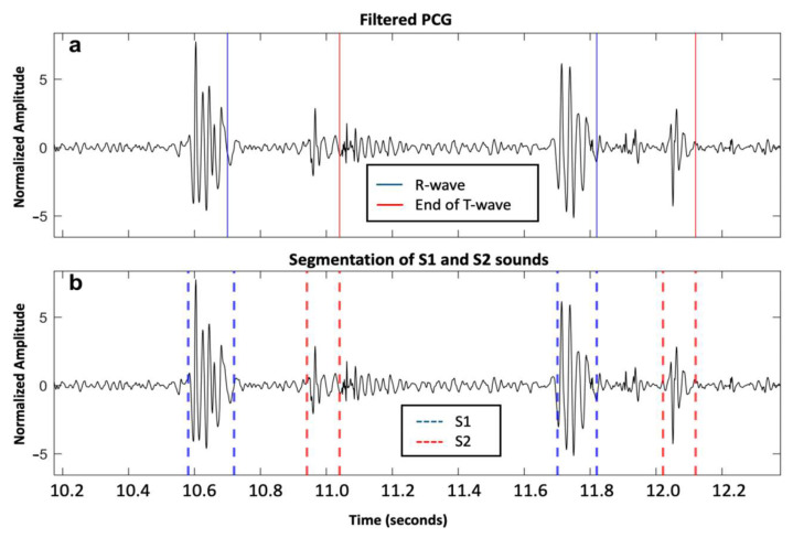
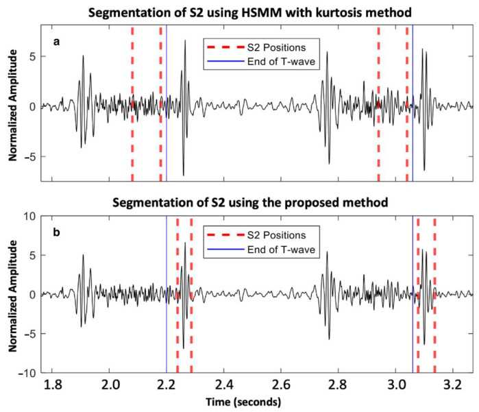
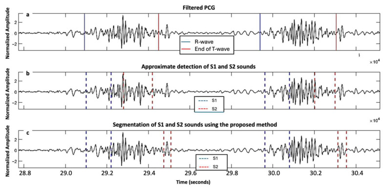
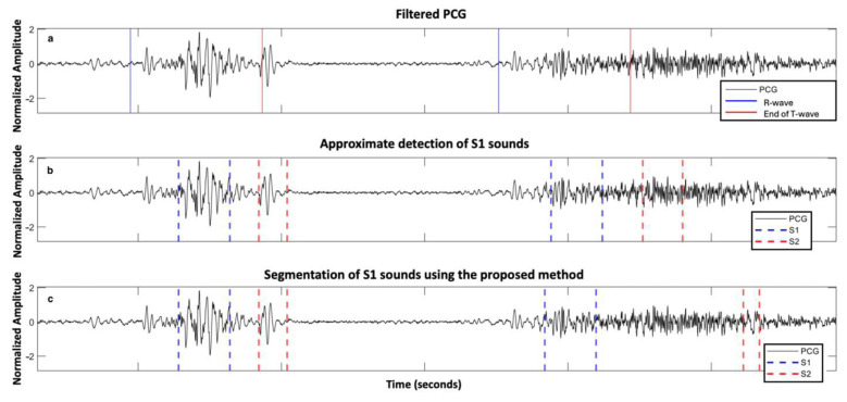
Severe aortic valve diseases (AVD) cause changes in heart sounds, making phonocardiogram (PCG) analyses challenging. This study presents a novel method for segmenting heart sounds without relying on an electrocardiogram (ECG), specifically targeting patients with severe AVD. Our algorithm enhances traditional Hidden Semi-Markov Models by incorporating signal envelope calculations and statistical tests to improve the detection of the first and second heart sounds (S1 and S2). We evaluated the method on the PhysioNet/CinC 2016 Challenge dataset and a newly acquired AVD-specific dataset. The method was tested on a total of 27,400 cardiac cycles. The proposed approach outperformed the existing methods, achieving a higher sensitivity and positive predictive value for S2, especially in the presence of severe heart murmurs. Notably, in patients with severe aortic stenosis, our proposed ECG-free method improved S2 sensitivity from 41% to 70%.

Keywords: aortic regurgitation; aortic stenosis; automatic detection; phonocardiography; telemedicine; valvular heart disease.


Conflict of interest statement

The authors declare no conflicts of interest.

Figures

Figure 1
Figure 1
Workflow of the proposed method for ECG-free heart sound segmentation, divided into two main phases: training and testing. In the training phase, ECG and PCG signals from a training dataset undergo R-peak and T-wave detections (ECG), as well as feature extraction (PCG). These features are used for heart sound labeling, which facilitates the training of a Hidden Semi-Markov Model (HSMM). The trained HSMM model is then applied in the testing phase, where ECG is only used for performance evaluation. Initial positions of the first and second fundamental heart sounds (S1 and S2) are identified, followed by wavelet envelope analysis and Shannon energy calculation to refine these positions. The final S1 and S2 positions are determined and evaluated for segmentation performance.
Figure 2
Figure 2
Two cardiac cycles on a phonocardiography (PCG) signal recorded from a healthy individual from the PhysioNet/CinC Challenge 2016 dataset: (a) Filtered PCG signal with R-wave and end of T-wave reported as blue and red solid lines, respectively; and (b) segmentation of first and second fundamental heart sounds (S1 and S2) reported as blue and red dashed lines, respectively, based on the proposed algorithm.
Figure 3
Figure 3
Aortic valve disease subject from the PhysioNet/CinC Challenge 2016 dataset: (a) segmentation of S2 using HSMM with kurtosis method [14]; and (b) segmentation of S2 using the proposed method.
Figure 4
Figure 4
Signals acquired from one subject with severe aortic stenosis, mild aortic regurgitation, and mild mitral regurgitation from the ARTIK dataset: (a) filtered PCG with R-peak and end of T-wave annotated; (b) segmentation of first and second fundamental heart sounds (S1 and S2) based on the HSMM with kurtosis method; and (c) segmentation of S1 and S2 using the proposed approach.
Figure 5
Figure 5
Signals acquired from a second subject with severe aortic stenosis, mild aortic regurgitation and mild mitral regurgitation from the ARTIK dataset: (a) filtered PCG with R-peak and end of T-wave annotated; (b) segmentation of first and second fundamental heart sounds (S1 and S2) based on the HSMM with kurtosis method; and (c) segmentation of S1 and S2 using the proposed approach.
Figure 6
Figure 6
Example of errors in segmenting two heartbeats in a subject with mixed aortic valve disease with predominant severe aortic stenosis from the ARTIK dataset: (a) PCG filtered with R-peaks and end of T-waves reported; (b) approximate segmentation of S1 and S2 sound; and (c) segmentation of S1 and S2 sound using the proposed approach.
Figure 7
Figure 7
Mild aortic regurgitation patient from the ARTIK dataset: (a) filtered PCG with R-peaks and end of T-waves reported; (b) approximate segmentation of S1; and (c) accurate segmentation of S1 using the proposed approach.

References

    1. Timmis A., Vardas P., Townsend N., Torbica A., Katus H., De Smedt D., Gale C.P., Maggioni A.P., Petersen S.E., Huculeci R., et al. European Society of Cardiology: Cardiovascular disease statistics 2021. Eur. Heart J. 2022;43:716–799. doi: 10.1093/eurheartj/ehab892. - DOI - PubMed
    1. Généreux P., Schwartz A., Oldemeyer J.B., Pibarot P., Cohen D.J., Blanke P., Lindman B.R., Babaliaros V., Fearon W.F., Daniels D.V., et al. Transcatheter Aortic-Valve Replacement for Asymptomatic Severe Aortic Stenosis. N. Engl. J. Med. 2024;392:217–227. doi: 10.1056/NEJMoa2405880. - DOI - PubMed
    1. Iung B. A worldwide perspective on the temporal burden and impact of calcific aortic valve disease. Eur. Heart J. Qual. Care Clin. Outcomes. 2023;9:439–441. doi: 10.1093/ehjqcco/qcad026. - DOI - PubMed
    1. Zhang N.S., Yang J.Y., Goldhaber J.I., Phan B.A.P., Cheitlin M.D. Cardiac auscultation skills among medical trainees. Am. Heart J. 2025;286:14–17. doi: 10.1016/j.ahj.2025.03.006. - DOI - PMC - PubMed
    1. Thoenes M., Bramlage P., Zamorano P., Messika-Zeitoun D., Wendt D., Kasel M., Kurucova J., Steeds R.P. Patient screening for early detection of aortic stenosis (AS)—Review of current practice and future perspectives. J. Thorac. Dis. 2018;10:5584–5594. doi: 10.21037/jtd.2018.09.02. - DOI - PMC - PubMed

LinkOut - more resources