. 2025 Sep;13(18):e70520.
doi: 10.14814/phy2.70520.

Spinal excitability following sensory electrical stimulation of the upper limb

Affiliations

Spinal excitability following sensory electrical stimulation of the upper limb

Devin Box et al. Physiol Rep. 2025 Sep.

Abstract

The purpose of the study was to determine if sensory electrical stimulation (SES), below motor threshold, would reduce spinal excitability via reciprocal inhibition (RI) and determine if any changes were sex-related. Eighteen healthy participants (11 males and 7 females) participated in a pre-post comparison study. The Hoffmann (H-) reflex was elicited to assess the spinal excitability of Flexor Carpi Radialis (FCR) and the influence of RI from Extensor Carpi Radialis Longus (ECRL) on FCR using a paired conditioning pulse paradigm. A 15-min bout of SES (4 pulse bursts at 100 Hz) was applied to ECRL, and the H-reflexes were measured at 0- and 20-min post SES. A linear mixed model regression analysis was performed to evaluate the effects of stimulus order, conditioning, sex, and time on the FCR H-reflex. All participants experienced RI from the conditioning pulses, with females having significantly greater suppression than males (mean difference; MD = 0.026). For males, SES produced a depression in FCR excitability (MD = 0.023 at time 0; MD = 0.015 at 20 min post-SES) with no changes in RI. SES had no effect on FCR excitability or RI in females. The potential for SES to produce changes in antagonist excitability was sex-related, which may have important rehabilitation considerations.

Keywords: reciprocal inhibition; spinal excitability; upper limb.


Conflict of interest statement

The authors have no real or perceived conflicts of interest to disclose relating to the work presented herein.

Figures

FIGURE 1
FIGURE 1
Protocol timeline. Determination of the intensity of test stimuli: H‐M recruitment curve from the flexor carpi radialis (FCR) muscle to find the intensity for the test H‐reflex. Maximal M‐wave from the extensor carpi radialis longus (ECRL) muscle to determine conditioning stimuli intensity. Reciprocal inhibition (RI) measures (striped rectangles) were taken before sensory electrical stimulation (SES) applied for 15 min (Pre), immediately after the SES (Post) and 20 min after SES (Post20).
FIGURE 2
FIGURE 2
(a) Schematic of the testing setup. Participants laid supine on a testing bed with their right arm abducted at a 45‐degree angle. The right arm was supported by an apparatus on an adjacent testing table. The gap between the two testing tables provided direct access to the stimulating sites on the upper arm. (b and c) Diagrams of recording and stimulating sites on the anterior aspect of the right arm (b), depicting the electrodes used for median nerve stimulation and flexor carpi radialis (FCR) EMG recording, and the posterior aspect of the right arm (c), depicting the electrodes used for radial nerve and extensor carpi radialis longus (ECRL) muscle stimulation, and ECRL EMG recording.
FIGURE 3
FIGURE 3
H‐M recruitment curve from a representative participant. Stimulation intensity required for eliciting a maximal H‐reflex (H‐max) and a maximal M‐wave (M‐max) was identified.
FIGURE 4
FIGURE 4
Flexor carpi radialis (FCR) and extensor carpi radialis longus (ECRL) maximal M‐waves from a single participant. (a) FCR M‐max before sensory electrical stimulation (SES) (Pre; solid black line), after SES (Post; dotted black line) and 20 min after SES (Post20; gray line). (b) Maximal ECRL M‐wave, obtained at the beginning of the experiment (thicker black line), used to find the stimulation intensity for conditioning stimulus (approximately 10% of M‐max). The antagonist M‐wave (expanded, right) elicited by the conditioning stimulus did not change in amplitude during Pre (solid black line), Post (dotted black line) or Post20 (gray line).
FIGURE 5
FIGURE 5
Unconditioned and conditioned H‐reflexes from a female (a) and a male (b) participant. Note the larger change in peak‐to‐peak amplitude in the conditioned H‐reflex in the female participant.
FIGURE 6
FIGURE 6
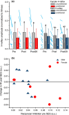
(a) Unconditioned and conditioned H‐reflexes at Pre, Post, Post20 for female (left) and male (right) participants. In both panels the average for each participant (Unconditioned to Conditioned) at each time point is shown with blue lines. The bars represent the mean with 95% confidence intervals for unconditioned (light gray—females; dark gray—males) and conditioned (red—females; dark blue—males) H‐ reflexes. * Denotes statistically significant difference from conditioned H‐reflex; + denotes statistically significant difference from Pre testing. (b) The change in reciprocal inhibition (RI) plotted against the Pre RI (before sensory electrical stimulation (SES)) for female (red circles) and male (dark blue triangles) participants. The data for each participant are averages of RI and the change of RI calculated between the pairs of unconditioned and conditioned H‐reflexes pre and post SES. Values below the horizontal line indicate less RI post SES than Pre.

References

    1. Ansdell, P. , Brownstein, C. G. , Škarabot, J. , Hicks, K. M. , Howatson, G. , Thomas, K. , Hunter, S. K. , & Goodall, S. (2019). Sex differences in fatigability and recovery relative to the intensity–duration relationship. The Journal of Physiology, 597(23), 5577–5595. - PubMed
    1. Beery, A. K. , & Zucker, I. (2011). Sex bias in neuroscience and biomedical research. Neuroscience and Biobehavioral Reviews, 35(3), 565–572. - PMC - PubMed
    1. Bergquist, A. J. , Clair, J. M. , Lagerquist, O. , Mang, C. S. , Okuma, Y. , & Collins, D. F. (2011). Neuromuscular electrical stimulation: Implications of the electrically evoked sensory volley. European Journal of Applied Physiology, 111(10), 2409–2426. - PubMed
    1. Burke, D. , Hallett, M. , Fuhr, P. , & Pierrot‐Deseilligny, E. (1999). H reflexes from the tibial and median nerves. The international federation of clinical neurophysiology. Electroencephalography and Clinical Neurophysiology. Supplement, 52, 259–262. - PubMed
    1. Crone, C. , Hultborn, H. , Jespersen, B. , & Nielsen, J. (1987). Reciprocal Ia inhibition between ankle flexors and extensors in man. The Journal of Physiology, 389(1), 163–185. - PMC - PubMed

LinkOut - more resources