. 2025 Sep 20;16(1):200.
doi: 10.1186/s13244-025-02073-8.

A pictorial essay of thoracic wall diseases: multiple pathologies in the same anatomical site

Affiliations

A pictorial essay of thoracic wall diseases: multiple pathologies in the same anatomical site

Giuliana La Rosa et al. Insights Imaging. .

Abstract

Objectives: To describe diagnostic and radiological features of the main pathologies affecting the thoracic wall, providing a pictorial atlas based on several clinical cases extracted from our archive.

Materials and methods: A wide variety of pathologies affect the tissues of the thoracic cage; these conditions are often encountered by radiologists during examinations performed for unrelated clinical questions. Modern imaging techniques enable the detection of these pathologies and allow definitive diagnoses to be achieved.

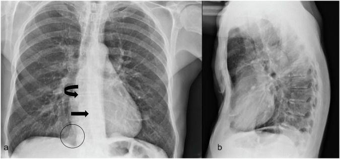
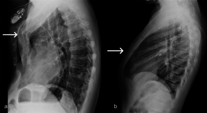
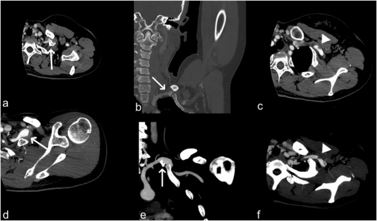
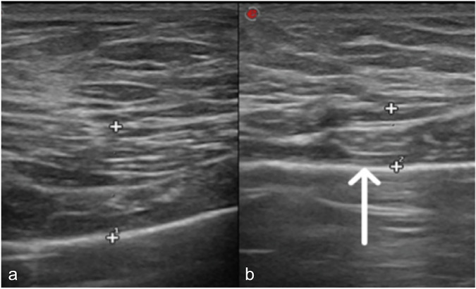
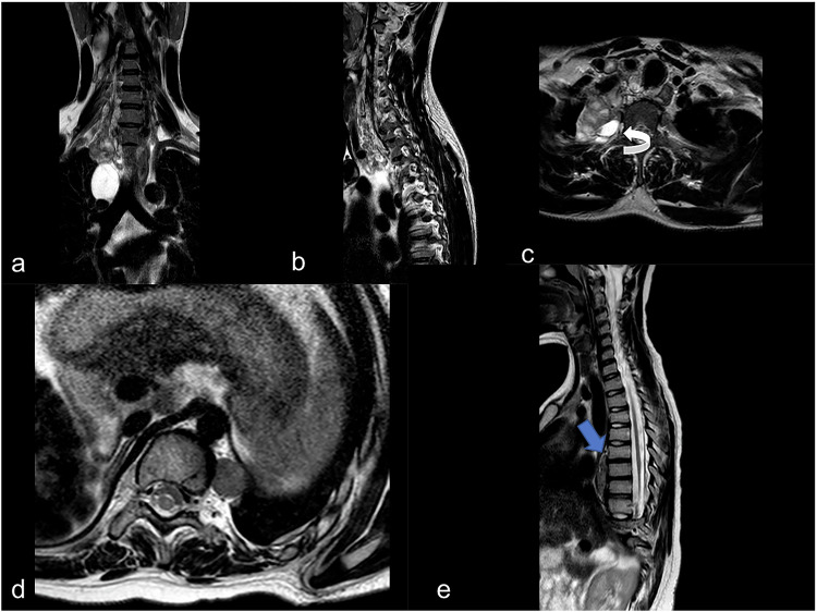
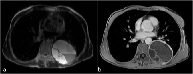
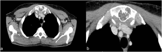
Results: Pathological processes that involve the chest wall may be classified into: (1) congenital and developmental diseases: pectus excavatum, pectus carinatum, supernumerary rib syndrome, Poland syndrome, neurofibromatosis, osteogenesis imperfecta, mucopolysaccharidosis, Marfan syndrome; (2) infectious and inflammatory diseases-such as aspergillosis, tuberculosis, abscesses from pyogenic bacteria, Tietze's syndrome; (3) bone injuries (traumatic and degenerative diseases): sternal, vertebral and costal fractures, degenerative disc and arthrosis pathology; (4) chest wall tumors-such as sarcomas, lymphomas, neurogenic tumors, lipoma.

Conclusions: Thoracic wall pathologies include a wide spectrum of conditions, with some clinical implications that often require a correct nosological framing. Recognizing these pathologies is essential for radiologists so that they can make a correct description in the report and direct toward appropriate treatment if required.

Critical relevance statement: Cage diseases are various and difficult to understand, so multimodality imaging plays a crucial role in achieving an efficient and final diagnosis.

Key points: Thoracic wall pathologies have different etiologies. Imaging represents a fundamental tool to clarify their extension, location, and nature. The prognosis of some of these diseases can be poor.

Keywords: Infections; Magnetic resonance imaging; Multidetector computed tomography; Neoplasms; Thoracic wall.


Conflict of interest statement

Declarations. Ethics approval and consent to participate: Not applicable. Consent for publication: Anonymized use of these images for research and publication is covered by institutional policy. Competing interests: S.P. is a member of the scientific editorial board of Insights into Imaging (section: Abdominal) and, as such, did not participate in the selection or review processes for this article. The remaining authors declare that they have no competing interests related to this article.

Figures

Fig. 1
Fig. 1
Chest X-ray was performed in orthostatism in two standard projections. This is a case of a 61-year-old male patient with a history of cardiac arrythmia. The posteroanterior projection (a) shows increased inferiorly-medial right hypodiaphany, related to increased visibility of the parasternal soft tissues located in the anterior chest wall (black circle), blurring of the right cardiac margin (curved black arrow) and axial rotation of heart with dislocation to the left side in the corresponding area of descending aorta (black arrow); on lateral view, the degree of depression could be easily assessed (b)
Fig. 2
Fig. 2
Pectus carinatum. Lateral chest X-ray views demonstrate the two types of sternal protrusion: chondromanubrial with protrusion of the manubrium and upper sternal portion (a), and chondrogladiolar with protrusion of the median and inferior region of the sternum (b)
Fig. 3
Fig. 3
A 35-year-old female patient with a supernumerary rib. CT images after contrast medium injection, in arterial phases (ad) and the same phase after maximum intensity projections reconstruction (e, f). They show the supernumerary rib, which causes the development of a thoracic outlet syndrome (white arrows in a—axial view and soft tissue window, and b—coronal view with bone window). c and f also demonstrate partial thrombosis of the subclavian artery (white arrowheads)
Fig. 4
Fig. 4
A 12-year-old male affected by Poland syndrome. Ultrasound images reveal normal pectoral muscle on the right side (a) and left pectoral muscle atrophy (white arrow in b)
Fig. 5
Fig. 5
X-ray examination, spine and thorax studies. A pediatric patient with mental and developmental illness, suffering from mucopolysaccharidoses. a A scoliotic attitude of the thoracolumbar spine is well depicted (white arrow). Widened and extended-looking ribs, especially the lower one, could be observed in a and b (curved arrow in b). Vertebral bodies are rounded and flattened (white arrowhead)
Fig. 6
Fig. 6
Patient affected by osteogenesis imperfecta. X-ray pictures show diffuse alteration of the vertebral bodies with a reduction in height (platyspondyly) and depression of the upper side of vertebral bodies—namely at the thoracic level (white arrow, b); marked gibbous deformation of the dorsal spine (b). Mildly dysmorphic appearance of the rib cage (a). MRI shows an area of signal hyperintensity in T2/STIR of the soma of Th6 due to edema (possible hyperemia or microfracture) (c, white circle). The intervertebral discs take an oval appearance in the absence of significant protrusions (c, white arrowhead)
Fig. 7
Fig. 7
Typical case of Tietze syndrome (CT images, bone view): young man patient 30 years old, with costal pain and no history of inflammatory condition, autoimmune or neoplastic condition. Tietze syndrome is a benign condition characterized by an inflammation of the costosternal cartilage, often with hypertrophy. a Axial CT image (bone window) shows focal swelling and sclerosis of the second costosternal joint, with surrounding soft tissue thickening. b Coronal reformatted CT image confirms enlargement and sclerosis of the costal cartilage at the same level, consistent with Tietze syndrome
Fig. 8
Fig. 8
Rib chondrosarcoma in a 30-year-old male patient. a Axial contrast-enhanced CT image (venous phase) shows a soft tissue mass originating from the left costal cartilage with adjacent chondral erosion (white arrow), without extension into the underlying muscle layers. b Sagittal reconstruction confirms the cartilaginous origin and the absence of deep muscle invasion
Fig. 9
Fig. 9
MRI-T2 sequences with fat suppression. ac Neurofibroma arising from the neck and extending to the upper thoracic site, (c) widened right neural exit foramina (white curved arrow). d, e A typical case of pediatric paravertebral neurinoma with a rounded aspect (d). In this case, lesions do not cause bone erosion but vertebral scalloping in the thoracic vertebral body at the level of Th5 (blue arrow). Both tumors show shade hyperintense aspects on T2-weighted sequences
Fig. 10
Fig. 10
Magnetic resonance is a promising imaging modality to confirm neurogenic tumors (like paragangliomas)—the internal fluid produces a hyperintense signal on T2-weighted MRI images (a). b A typical hypointense aspect on the T1-weighted image
Fig. 11
Fig. 11
Secondary rib lesions from hepatocellular carcinoma in a patient with known liver malignancy. a Coronal CT image in the basal phase shows an oval-shaped, expansile lesion invading the rib. b Axial CT image in the arterial phase demonstrates the hypervascularization of the lesion, consistent with secondary spread of hepatocellular carcinoma
Fig. 12
Fig. 12
Sternal enchondroma in a 66-year-old male patient. a Axial CT image in mediastinal window shows a lytic lesion with defined margins involving the manubrium sterni. b More cranial axial CT slice confirms the cartilaginous nature of the lesion, with absence of aggressive features such as soft tissue extension or cortical disruption

References

    1. Dähnert W (2007) Radiology review manual 6th ed. Lippincott Williams & Wilkins, Philadelphia, PA
    1. Kelly RE, Cash TF, Shamberger RC et al (2008) Surgical repair of pectus excavatum markedly improves body image and perceived ability for physical activity: multicenter study. Pediatrics 122:1218–1222 - PubMed
    1. Garcia VF, Seyfer AE, Graeber GM (1989) Reconstruction of congenital chest-wall deformities. Surg Clin North Am 69:1103–1118 - PubMed
    1. Takahashi K, Sugimoto H, Ohsawa T (1992) Obliteration of the descending aortic interface in pectus excavatum: correlation with clockwise rotation of the heart. Radiology 182:825–828 - PubMed
    1. Haller JA, Kramer SS, Lietman SA (1987) Use of CT scans in selection of patients for pectus excavatum surgery: a preliminary report. J Pediatr Surg 22:904–906 - PubMed

LinkOut - more resources