. 2025 Sep 22;10(1):313.
doi: 10.1038/s41392-025-02390-w.

Spatiotemporal multi-omics analysis uncovers NAD-dependent immunosuppressive niche triggering early gastric cancer

Affiliations

Spatiotemporal multi-omics analysis uncovers NAD-dependent immunosuppressive niche triggering early gastric cancer

Pingting Gao et al. Signal Transduct Target Ther. .

Abstract

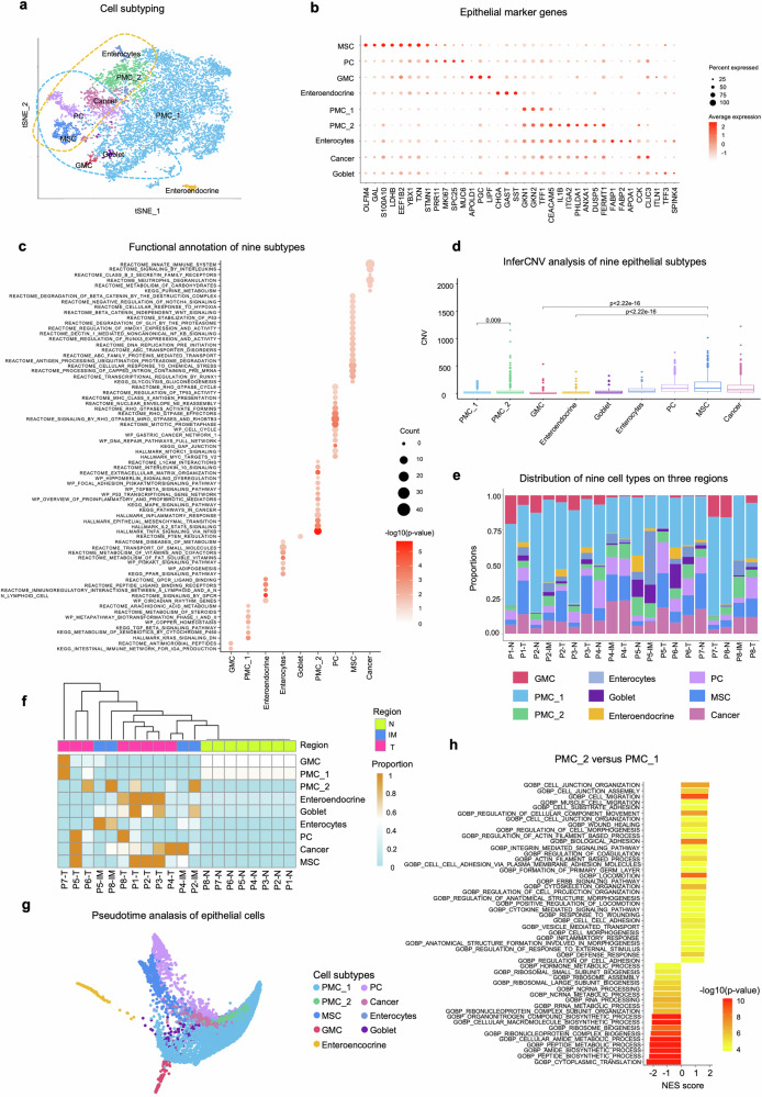
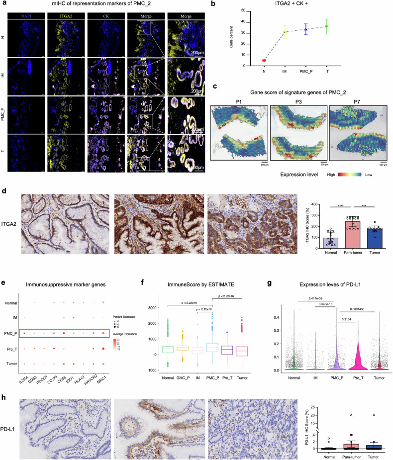
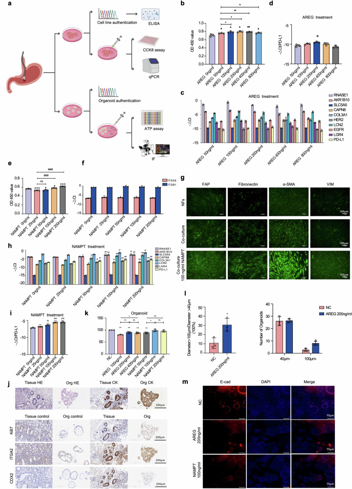
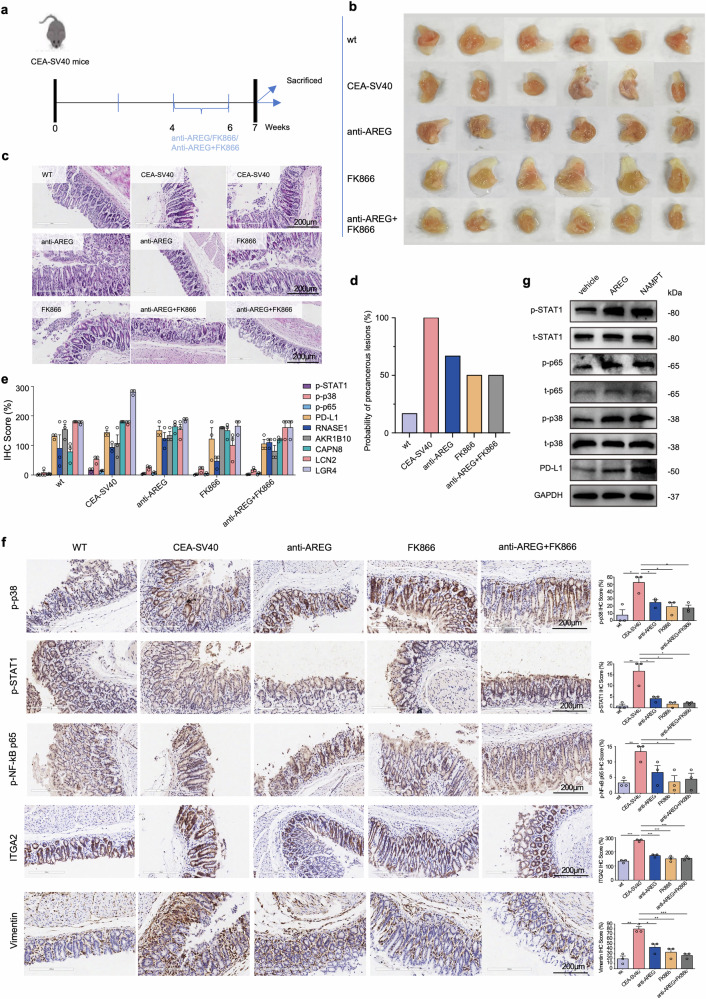
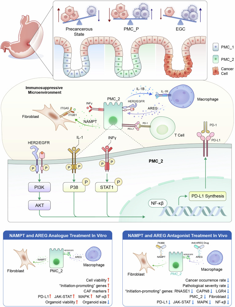
Understanding the cellular origins and early evolutionary dynamics that drive the initiation of carcinogenesis is critical to advancing early detection and prevention strategies. By characterizing key molecular, cellular and niche events at the precancerous tipping point of early gastric cancer (EGC), we aimed to develop more precise screening tools and design targeted interventions to prevent malignant transformation at this stage. We utilized our AI models to integrate spatial multimodal data from nine EGC endoscopic submucosal dissection (ESD) samples (covering sequential stages from normal to cancer), construct a spatial-temporal profile of disease progression, and identify a critical tipping point (PMC_P) characterized by an immune-suppressive microenvironment during early cancer development. At this stage, inflammatory pit mucous cells with stemness (PMC_2) interact with fibroblasts via NAMPT ITGA5/ITGB1 and with macrophages via AREG EGFR/ERBB2 signaling, fostering cancer initiation. We established gastric precancerous cell lines and organoids to demonstrate that NAMPT and AREG promote cellular proliferation in vitro. Furthermore, in the transgenic CEA-SV40 mouse model, targeting AREG and/or NAMPT disrupted key cell interactions, inhibited the JAK-STAT, MAPK, and NFκB pathways, and reduced PD-L1 expression, which was also confirmed by western blot in vitro. These interventions delayed disease progression, reversed the immunosuppressive microenvironment, and prevented malignant transformation. Clinical validation was conducted using endoscopically resected EGC specimens. Our study provides a precise spatiotemporal depiction of EGC development and identifies novel diagnostic markers and therapeutic targets for early intervention.


Conflict of interest statement

Competing interests: The authors declare no competing interests.

Figures

Fig. 1
Fig. 1
Spatial-temporal heterogeneity of early gastric lesions encompassing normal, intestinal metaplasia (IM), and tumor (T) regions. a Schematic overview of the experimental workflow. Fresh ESD samples containing adjacent normal (N), intestinal metaplasia (IM), and tumor (T) tissues were obtained from nine patients. b UMAP visualization of single-cell RNA sequencing (scRNA-seq) data showing 12 major cell types. c Dot plot illustrating the expression of canonical marker genes across identified cell types. Dot size represents the percentage of cells expressing each gene; color scale indicates the average normalized expression level. d H&E-stained spatial transcriptomics (ST) section from patient P1, with histologically annotated normal (N), early tumor (ET), and tumor (T) regions. e Spatial domains identified by the stMVC algorithm based on ST data from patient P1. f UMAP visualization of spatial domains identified by stMVC in ST data of patient P1. g The bar chart showing the proportion of 12 cell types identified from scRNA-seq across each histological in the ST data from eight patients. h The heatmap displaying the enrichment of 12 cell types identified from scRNA-seq across 20 histologically annotated regions in the ST data from eight patients
Fig. 2
Fig. 2
Heterogeneity of epithelial cell subtypes. a tSNE plot illustrating nine epithelial cell subtypes identified from scRNA-seq data. b Dot plot showing the expression levels of marker genes in each subtype. Dot size and color indicate the percentage and mean expression level of each gene. c Functional annotation of nine subtypes. d Boxplot displaying large-scale copy number variations (CNVs) in six subtypes (goblet, enterocytes, PMC_2, PC, MSC, and cancer) compared to PMC_1, GMC, and enteroendocrine subtypes. Each color indicates one subtype. e Distribution of the nine cell subtypes across N, IM, and T regions in the ST data from each patient. f Unsupervised clustering analysis based on the proportions of epithelial subtypes across normal, IM, and cancer regions. g Pseudotime trajectory analysis of epithelial cells derived from scRNA-seq data from all single-cell samples. h Functional analysis comparing PMC_2 to PMC_1
Fig. 3
Fig. 3
Identification of critical epithelial cell states driving EGC initiation. a Dot plot showing the expression of representative marker genes across six spatial epithelial groups identified in ST data. Dot size indicates the percentage of cells expressing each gene; color scale denotes mean normalized expression level. b Box plot showing large-scale CNV levels across six spatial groups. CNV levels were normalized across samples; each color represents a different group. c Gene signature scores of GMC_P (top) and PMC_P (bottom) marker genes across ST regions in three representative patients (P1, P2, and P3). d Overall survival rate of patients based on the average expression of seven signature genes of PMC_P in stomach cancer from the TCGA database by GEPIA2. e Gene set enrichment analysis (GSEA) of differentially expressed genes in PMC_P versus IM. f Heatmap showing the proportion of nine epithelial subtypes in six groups of ST data
Fig. 4
Fig. 4
Precancerous PMC_P niche coordinates PMC_2 accumulation and PD-L1-mediated immune suppression. a Spatial distribution of PMC_2 markers (CK/ITGA2) across these groups via immunofluorescence double labeling (DAPI/CK/ITGA2). Co-localization signals (Merge, white) of CK (purple) and ITGA2 (yellow) show enrichment of PMC_2 cells in the PMC_P region indicating their expansion starting from the precancerous microenvironment. b Quantitative analysis further confirms a gradient increase in PMC_2 proportion across progression stages (N < IM < PMC_P < T). c Spatial expression of the gene signature score of PMC_2 in three representative patients: P1, P3, and P7. d ITGA2 expression in 21 EGC patients showed significant higher expression level of ITGA2 in para-tumor regions than other regions. Scale bar: 200 μm. e Dot plot showing the expression of classical immunosuppressive marker genes, with IL2RA, CD33, PDCD, CD274, CD86, IDO1, HLA-G, HAVCR2, and MRC1 highly expressed in PMC_P region. Dot size indicates detection rate, color scale represents normalized expression. f Boxplot of the distribution of immune score (calculated by ESTIMATE) across the six groups showing significantly higher immune score in PMC_P region than normal or tumor. g Violin plot showing the expression level of PD-L1 across six groups which is significantly higher in PMC_P region. h PD-L1 expression in 21 EGC patients showing higher expression of PD-L1 in para-tumor regions than other regions. Scale bar: 200 μm. ***p < 0.001, ****p < 0.0001
Fig. 5
Fig. 5
Identification of key cell populations and CCC mechanisms in the initiation of EGC. a Scatter plot showing the specificity (Gini index) and difference level (logPvalue) of over-expressed ligand genes in the PMC_P group. b Bubble heatmap displaying mean CCC strength by CellChat for AREG, IL1B, NAMPT, KLK7, IGFBP3, PRSS3, CXCL5, and POSTN interaction pairs across different domains in three representative patients: P1, P3, and P7. c, d Spatial expression of the ligand AREG, receptors EGFR and Erb-B2 receptor tyrosine kinase 2 (ERBB2), and their corresponding CCC interactions in three representative patients: P1, P3, and P7. e H&E plot with four groups: N, IM, PMC_P, and T in the upper panel with a scale bar of 200 µm. mIHC images of the four groups for AREG (green), EGFR (yellow), and ERBB2 (red), with a scale bar of 20 µm showed over-expression of these three genes in the PMC_P. Red arrows: AREG+ cells and HER2 + EGFR+ cells co-localization. f H&E plot with four groups: N, IM, PMC_P, and T in the upper panel with a scale bar of 1 mm. mIHC images of the four groups for NAMPT (green), ITGA5 (yellow), and ITGB1 (red), with a scale bar of 20 µm, showed over-expression of these three genes in the PMC_P. Red arrows: ITGB1 + ITGA5+cells and NAMPT+ cells co-localization
Fig. 6
Fig. 6
PMC_2 interacts strongly with fibroblasts and macrophages within PMC_P. a, b Circle plot (based on pan-tissue scRNA-seq across regions) displaying the intercellular communication between different cell types for AREG EGFR/ERBB2, and NAMPT ITGA5/ITGB1. c Spearman correlation (region-specific analysis-PMC_P only) between AREG expression and gene signature scores for enterocytes (FABP1, FABP2, and APOA1), PMC_2 (CEACAM5, ITGA2, PHLDA1, ANXA1, DUSP5, and FERMT1), NKT cells (CD160, KRT81, and KIR2DL4), mast cells (TPSB2, MS4A2, TPSAB1, and KIT), and myeloid cells (S100A8, S100A9, IL1B, and CD68) (left), NAMPT expression and gene signature scores for PMC_2 (CEACAM5, ITGA2, PHLDA1, ANXA1, DUSP5, and FERMT1), neutrophil (FCGR3B, CXCR2, IFITM2, CMTM2, and CSF3R), mDC (PPP1R14A, FCER1A, HLA-DPB1, and CD1C), CD14 mono (GIMAP7, AHNAK, and CD36), monocyte (CCDC88C, and POU2F2), M2 macrophage (SLCO2B1, C1QC, C1QA, GPNMB, and C1QB), and VEGFA + M1 (TGM2, CLEC5A, VEGFA, and MCEMP1) (right). d mIHC staining shows ligand-receptor pair NAMPT (secreted by PMC_2 marked by CK + ITGA2+) ITGA5/ITGB1 (expressed by fibroblasts marked by DCN) across four regions: N, IM, PMC_P, T. The magnified view within the boxed region highlights NAMPT + PMC_2 cells (white arrows) and ITGB1 + ITGA5+fibroblast cells (red arrows). Scale bar: 50 μm. e mIHC staining illustrates ligand-receptor pair AREG (expressed by macrophages marked by S100A8) EGFR/ERBB2 (expressed by PMC_2 marked by CK, and ITGA2) across four regions: N, IM, PMC_P, T. The magnified view within the boxed region highlights EGFR + ERBB2 + PMC_2 cells (white arrows) and AREG+ macrophages (red arrows). Scale bar: 50 μm. f Line charts showing the proportion of AREG+ cells, EGFR + ERBB2 + PMC_2 cells, ITGA5 + ITGB1+ fibroblasts, NAMPT + PMC_2 cells across four regions: N, IM, PMC_P, T. g Distribution of four myeloid cell subtypes showing macrophage as main myeloid cell type in PMC_P region. h Spearman correlation between AREG and myeloid subclusters: mDC (PPP1R14A, FCER1A, HLA-DPB1, and CD1C), CD14 mono (GIMAP7, AHNAK, and CD36), M2 macrophage (SLCO2B1, C1QC, C1QA, GPNMB, and C1QB), and VEGFA + M1 (TGM2, CLEC5A, VEGFA, and MCEMP1). i Spatial distribution of three histological status (i.e., N, IM, and T), spatial clusters by stMVC (N, IM, PMC_P, and T), distributions of five cell types, and zoomed-in views of the cell type distributions from PMC_P to T
Fig. 7
Fig. 7
AREG and NAMPT enhance proliferation and gene modulation in precancerous gastric cell lines and organoids. a Precancerous cell lines (Pre-EGC) and organoids were successfully established from biopsies of EGC patients and treated with AREG and/or NAMPT. b CCK-8 assay demonstrating the impact of AREG on the viability of Pre-EGC after 72 h. c qPCR analysis of Pre-EGC treated with AREG for 72 h showing changes in the expression of “initiation-promoting” genes, HER2, EGFR and PD-L1. d qPCR analysis highlighting dose-dependent modulation of PD-L1 expression in Pre-EGC following AREG treatment (with 200 ng/ml the highest). e CCK-8 assay demonstrating the impact of NAMPT (0, 25, 50, 100, 200 ng/ml) on the viability of Pre-EGC after 72 h, with significant proliferation observed at 100 ng/ml. f Dose-dependent upregulated expressions of fibroblast surface receptors ITGA5 and ITGB1, with the most significant increases observed at 25 ng/ml and 50 ng/ml, respectively. g Enhanced fluorescence intensity of CAF markers (FAP, fibronectin, α-SMA and VIM) observed in NFs co-cultured with Pre-EGC treated with 100 ng/ml NAMPT compared to normal fibroblasts (NFs) and untreated groups. h PCR analysis of Pre-EGC co-cultured with NFs treated with NAMPT for 72 h showing changes in the expression of “initiation-promoting” genes and PD-L1. i qPCR analysis highlighting dose-dependent modulation of PD-L1 expression in Pre-EGC following NAMPT treatment (significant with 100 ng/ml). j Validation and morphological characterization of precancerous gastric organoids. k ATP assay illustrating the proliferative effects of AREG on precancerous organoids after 5 days of treatment. l Organoids treated with 200 ng/ml AREG for 5 days were analyzed for size and gene expression. The left panel shows the number of organoids with diameters greater than 100 µm in the treatment group compared to controls. The right panel shows the proportion of organoids larger than 100 µm/40 µm in the treated group compared to controls. m Immunofluorescence analysis of E-cadherin in organoids treated with 200 ng/ml AREG or 100 ng/ml NAMPT for 5 days, revealing differential expression patterns. *p < 0.05, **p < 0.01, ***p < 0.01; #p < 0.05, ##p < 0.01, ###p < 0.001; ns no significance
Fig. 8
Fig. 8
Therapeutic efficacy of anti-AREG and FK866 in the CEA-SV40 mouse model of EGC. a Schematic flowchart outlining the induction of EGC in CEA-SV40 mice and the oral administration protocol for anti-AREG, FK866, and the combination treatment (anti-AREG + FK866). b Representative images of mouse stomachs dissected longitudinally to expose the gastric mucosa for macroscopic examination. c Macroscopic examination of early lesions revealed no visible abnormalities in all groups. However, H&E staining identified distinct histological features: deep nuclear staining and polarity alterations were prominent in the untreated CEA-SV40 group, while the anti-AREG group exhibited mild dysplasia, and the FK866 and combined treatment groups showed the least dysplasia. 200 μm. d Probability of precancerous lesion formation (%) across groups. The untreated CEA-SV40 mice showed the highest incidence (~100%), while anti-AREG and FK866 treatments significantly reduced lesion probability. The combined treatment group demonstrated an approximately 50% reduction, comparable to individual treatments. e Immunohistochemical staining scores for “initiation-promoting” gene-set, NFκB, JAK/STAT, and MAPK pathways showed distinct expression patterns across groups. f Representative IHC staining images for PMC_2 markers (ITGA2), fibroblast marker (Vimentin) along with key pathway markers (pp38, p-STAT1 and pp65) revealed significant differences among groups. g Western blot analysis of PD-L1, STAT, p-STAT1, p38, p-p38, p65, and p-p65 after 72-h treatments in Pre-EGC, Pre-EGC + 200 ng/ml AREG, and Pre-EGC + fibroblasts groups treated with 100 ng/ml NAMPT/200 ng/ml AREG. *p < 0.05, **p < 0.01, ***p < 0.01
Fig. 9
Fig. 9
Graphical summary of molecular, cellular, and microenvironmental features defining the precancerous tipping point that drives EGC initiation. Using AI-integrated spatial multi-modal analysis of ESD specimens, we defined a precancerous tipping point niche (PMC_P) characterized by distinct molecular, cellular, and microenvironmental features. Within PMC_P, inflammatory pit mucous cells with stem-like properties (PMC_2) engage in pathological crosstalk with fibroblasts via NAMPTITGA5/ITGB1 and with macrophages via AREGEGFR/ERBB2 signaling. These interactions collectively rewire oncogenic pathways, establish an immunosuppressive niche, and drive malignant transformation. The pivotal role of NAMPT and AREG signaling in EGC initiation was further validated through in vitro and in vivo models, including patient-derived cell lines, organoids and CEA-SV40 transgenic mice

References

    1. Smyth, E. C. et al. Gastric cancer. Lancet396, 635–648 (2020).
    1. Tan, P. & Yeoh, K. G. Genetics and molecular pathogenesis of gastric adenocarcinoma. Gastroenterology149, 1153–1162.e3 (2015). - PubMed
    1. Correa, P. A human model of gastric carcinogenesis. Cancer Res.48, 3554–3560 (1988). - PubMed
    1. Sugano, K., Moss, S. F. & Kuipers, E. J. Gastric intestinal metaplasia: real culprit or innocent bystander as a precancerous condition for gastric cancer?. Gastroenterology165, 1352–1366.e1 (2023). - PubMed
    1. de Vries, A. C. et al. Gastric cancer risk in patients with premalignant gastric lesions: a nationwide cohort study in the Netherlands. Gastroenterology134, 945–952 (2008). - PubMed

Substances