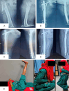
Short versus long proximal femoral nail anti-rotation-II (PFNA-II) in the management of unstable intertrochanteric fractures
- PMID: 40978687
- PMCID: PMC12444419
- DOI: 10.62347/LRTZ6852
Short versus long proximal femoral nail anti-rotation-II (PFNA-II) in the management of unstable intertrochanteric fractures
Abstract
Objectives: Unstable intertrochanteric (IT) fractures, particularly in elderly patients with low bone mineral density, pose significant treatment challenges. Proximal femoral nail anti-rotation-II (PFNA-II) is widely used, but the optimal implant length (short vs. long) remains debated. The objective of this study was to compare the clinical and functional outcomes of short versus long PFNA-II implants in unstable IT fractures.
Methods: A prospective comparative study was conducted at a tertiary hospital from November 2018 to November 2020. Adult patients (age ≥18) with recent (≤3 weeks) unstable IT femur fractures were included. Unstable fractures were defined by comminution of the posteromedial cortex, a compromised lateral wall (including reverse obliquity), or subtrochanteric extension. Patients with pathological fractures (other than osteoporosis), open fractures, polytrauma, pre-existing ipsilateral hip pathology, or non-ambulatory status were excluded. Patients were allocated to short PFNA-II (n=38) or long PFNA-II (n=40) groups based on the surgeon's intraoperative judgment (no randomization). All patients underwent standard reduction on a fracture table and fixation with PFNA-II. Postoperative mobilization and weight-bearing protocols were adjusted according to fracture stability and fixation quality. Outcome measures included fracture union time, complications, and the Harris Hip Score (HHS). Statistical significance was set at P<0.05.
Results: Both groups had similar demographics, fracture types, and surgical durations (P>0.05). Fracture union was achieved in 94.7% (36/38) of short-nail patients and 90% (36/40) of long-nail patients, with no significant difference in union rates or time to union (mean ~14 weeks, P>0.05). The short PFNA-II group demonstrated a significantly higher final HHS (87.2±7.1 vs. 82.3±7.8, P=0.03), with 89.5% achieving good/excellent outcomes vs. 62.5% in the long-nail group. Postoperative complications differed in pattern: anterior thigh pain was more frequent in short nails (15.8% vs. 2.5%), whereas mechanical complications (varus collapse >5°, helical blade lateral migration) were more common in long nails (15% vs. 5.3% varus collapse; 10% vs. 2.6% blade migration). However, overall complication rates were not significantly different between groups (P=0.17). No deep infections, implant breakage, or cut-out occurred in either group.
Conclusion: PFNA-II fixation is effective for unstable IT fractures with high union rates and low major complication rates in both implant groups. Short PFNA-II nails yielded superior functional outcomes and fewer mechanical complications compared to long nails in similar unstable fracture patterns. These findings suggest that implant length plays a crucial role in optimizing patient outcomes. In most cases of unstable IT fractures, a short PFNA-II appears advantageous, though patient anatomy (e.g. extreme femoral curvature) and fracture morphology should be considered when selecting implant length.
Keywords: Harris Hip Score; Intertrochanteric fracture (IT); NSA (neck shaft angle); implant length; proximal femoral nail anti-rotation-II (PFNA-II).
IJBT Copyright © 2025.
Conflict of interest statement
None.
Figures


References
-
- Cooper C, Campion G, Melton LJ 3rd. Hip fractures in the elderly: a world-wide projection. Osteoporos Int. 1992;2:285–9. - PubMed
-
- Gullberg B, Johnell O, Kanis JA. World-wide projections for hip fracture. Osteoporos Int. 1997;7:407–13. - PubMed
-
- Raja RSB, Singh D, Parasuraman K, Manikandarajan A. Management of unstable intertrochanteric fracture by proximal femoral nailing with trochanteric buttress plating. Int J Orthop Sci. 2022;8:47–52.
LinkOut - more resources
Full Text Sources
