Case Reports
. 2025 Aug 17;17(8):e90342.
doi: 10.7759/cureus.90342. eCollection 2025 Aug.

A Rare Case of Ewing Sarcoma of the Maxilla and a Literature Review of Similar Cases

Affiliations
Case Reports

A Rare Case of Ewing Sarcoma of the Maxilla and a Literature Review of Similar Cases

Abdul Moiz Khan et al. Cureus. .

Abstract

Ewing sarcoma (ES) is a small, round, blue cell malignant neoplasm that rarely occurs in the craniofacial skeleton, with presentation in the maxilla being exceedingly rare. We report the case of a 14-year-old girl with a two-year history of slowly progressive facial swelling that has recently become greatly enlarged. Imaging revealed an aggressive expansile left maxillary lesion with soft-tissue involvement and internal calcifications. Surgical resection was performed, and histopathological analysis showed sheets of homogenous round cells with minimal cytoplasm and round nuclei. Immunohistochemical staining for CD99, CD56, and NKX2.2 was positive, while staining for epithelial, muscle, and neural markers was negative, thereby confirming the diagnosis of ES. This case highlights the diagnostic difficulty of ES in unusual sites such as the maxilla, where it can mimic odontogenic or fibro-osseous lesions. Early diagnosis with histopathology and immunohistochemistry is important for appropriate management, which usually includes surgical resection followed by chemotherapy.

Keywords: anterior maxilla; ewing sarcoma (es); immunohistochemistry staining; pediatric bone tumor; small blue round cell tumor.


Conflict of interest statement

Human subjects: Informed consent for treatment and open access publication was obtained or waived by all participants in this study. Conflicts of interest: In compliance with the ICMJE uniform disclosure form, all authors declare the following: Payment/services info: All authors have declared that no financial support was received from any organization for the submitted work. Financial relationships: All authors have declared that they have no financial relationships at present or within the previous three years with any organizations that might have an interest in the submitted work. Other relationships: All authors have declared that there are no other relationships or activities that could appear to have influenced the submitted work.

Figures

Figure 1
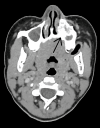
Figure 1. Axial CT of the face (soft-tissue window) demonstrating an expansile lytic maxillary mass (black arrows); biopsy-proven Ewing sarcoma in the (A) lower axial and (B) superior axial parts of the face.
(A) Lower axial: Expansile, lytic lesion centered in the left upper alveolar arch just superior to the first and second premolars (black arrow). The mass measures approximately 28.8 × 20.4 × 24.2 mm (AP × transverse × craniocaudal) and closely abuts the premolar roots, with focal cortical thinning/erosion. (B) More superior axial: The lesion contains internal punctate/coarse calcifications (black arrow) and displaces the unerupted left maxillary canine superiorly. Appearance reflects an aggressive, mineralizing tumor with local bone destruction, and biopsy confirmed Ewing sarcoma.
Figure 2
Figure 2. CT scan revealing no involvement of the left maxillary sinus by the tumor (black arrow).
Coronal CT scan illustrating that the lesion spares the left maxillary sinus (black arrow). Although the floor of the maxilla is thinned, there is no extension into the sinus cavity. The lesion extends anteriorly into the overlying skin and subcutaneous tissues and inferiorly into the gingival mucosa.
Figure 3
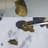
Figure 3. Gross resection specimen from the left maxilla (biopsy-proven Ewing sarcoma). (A) Close-up of tumor surface with adherent dentition; (B) specimen overview showing tooth-bearing fragments and tumor bulk.
(A) Close-up view of the resected specimen showing an irregular, lobulated tan-brown soft-tissue mass with adherent necrotic/hemorrhagic material and multiple attached teeth (visible crowns and roots), consistent with a tumor arising from and destroying the alveolus. (B) Overview of the specimen fragments demonstrating portions of the mass with embedded/extracted teeth and surrounding soft tissue; the specimen measured 5 × 4 × 3 cm on gross inspection.
Figure 4
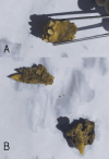
Figure 4. Cut sections of the resected left maxillary specimen (biopsy-proven Ewing sarcoma) demonstrating tumor infiltration of the alveolar bone with attached premolars and canines. (A) Close-up tooth-bearing fragment with tumor infiltration; (B) specimen fragments showing infiltrative cut surfaces and attached premolar/canine roots.
(A) Close-up of a larger tooth-bearing fragment: the cut surface is soft, tan-brown, and irregular, with a tumor infiltrating around and into the tooth roots (premolars/canine visible). (B) Additional smaller fragments showing infiltrative tumor tracking along root surfaces and into adjacent bone; one fragment demonstrates the pointed canine apex projecting from the tumor mass.
Figure 5
Figure 5. Malignant neoplasm showing round cells with scant cytoplasm and round nuclei with fragments of bone (black arrow).
Hematoxylin and eosin (H&E)-stained section (original magnification ×40) showing sheets of uniform small round blue cells with scant cytoplasm and round nuclei, separated by thin fibrous septa. Fragments of bone are visible (black arrow), indicating osseous invasion. The nuclear chromatin is finely stippled, and nucleoli are inconspicuous, features characteristic of Ewing sarcoma.
Figure 6
Figure 6. Cell showing positivity for CD99.
Immunohistochemistry (IHC) for CD99 showing diffuse, strong membranous positivity in tumor cells. CD99 expression is a characteristic but not entirely specific finding for Ewing sarcoma, aiding in diagnosis when interpreted in conjunction with morphology and other markers.
Figure 7
Figure 7. Cell showing positivity for CD56.
IHC for CD56 revealing strong cytoplasmic positivity in tumor cells. CD56 is a neural cell adhesion molecule that is often expressed in Ewing sarcoma, supporting the neuroectodermal origin of the tumor.
Figure 8
Figure 8. Cell showing positivity for NKX 2.2.
IHC for NKX2.2 showing nuclear positivity in tumor cells. NKX2.2 is a transcription factor expressed in the majority of Ewing sarcoma cases and, in combination with CD99 and CD56, supports the diagnosis and helps exclude histologic mimics.

References

    1. Management and outcome of Ewing sarcoma of the head and neck. Grevener K, Haveman LM, Ranft A, et al. Pediatr Blood Cancer. 2016;63:604–610. - PubMed
    1. Ewing sarcoma of the oral cavity. A review. Margaix-Muñoz M, Bagán J, Poveda-Roda R. J Clin Exp Dent. 2017;9:0–301. - PMC - PubMed
    1. Multimodality treatment in Ewing's sarcoma family tumors of the maxilla and maxillary sinus: review of the literature. Thorn D, Mamot C, Krasniqi F, Metternich F, Prestin S. Sarcoma. 2016;2016:3872768. - PMC - PubMed
    1. Ewing's sarcoma of the head and neck. Fonseca AS, Mezzalira R, Crespo AN, Bortoleto AE Jr, Paschoal JR. Sao Paulo Med J. 2000;118:198–200. - PMC - PubMed
    1. Ewing's sarcoma of maxillary sinus. Sharma R, Kumar P, Meher R, Shruti N, Gopal A, Mandal S. Indian J Otolaryngol Head Neck Surg. 2024;76:4760–4764. - PMC - PubMed

Publication types

LinkOut - more resources