. 2025 Sep 18;13(9):e70345.
doi: 10.1002/rcr2.70345. eCollection 2025 Sep.

Hemiclamshell Resection of a Ruptured Mediastinal Teratoma Exhibiting Pancreatic Differentiation

Affiliations

Hemiclamshell Resection of a Ruptured Mediastinal Teratoma Exhibiting Pancreatic Differentiation

Yuri Enomoto et al. Respirol Case Rep. .

Abstract

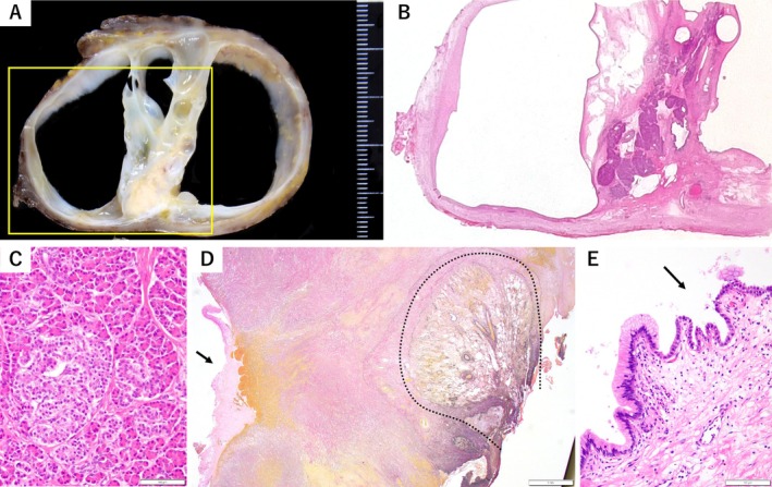
Mediastinal teratomas demonstrating unidirectional pancreatic differentiation are exceedingly rare. We describe a 36-year-old woman with progressively worsening anterior chest pain. Computed tomography demonstrated a 5.9 cm multilocular cystic mass in the anterior mediastinum, accompanied by pleural and pericardial effusions. Given the lesion's size and presumed inflammatory reaction, we performed radical excision via a hemiclamshell incision. Histopathology revealed predominantly pancreatic acinar tissue with scattered non-pancreatic epithelial elements, prompting reclassification as a mediastinal teratoma rather than ectopic pancreas. Focal epithelial disruption was regarded as the nidus of the surrounding inflammation. The patient's postoperative course was uneventful. This report underscores the need to include teratoma in the differential diagnosis of anterior mediastinal lesions initially interpreted as ectopic pancreas and illustrates the value of the hemiclamshell approach for achieving complete, atraumatic resection when extensive local adhesions are present.

Keywords: hemiclamshell approach; islets of Langerhans; mediastinal teratoma; pancreas; surgical resection.


Conflict of interest statement

The authors declare no conflicts of interest.

Figures

FIGURE 1
FIGURE 1
Radiological findings of the anterior mediastinal mass. (A) Chest radiograph revealing a new anterior mediastinal mass that was not present on imaging 2 years earlier. (B) Contrast‐enhanced CT showing a 5.9 × 6.1 × 6.8 cm anterior mediastinal mass, primarily composed of multilocular cystic components with minimal solid portions. Partial enhancement is seen along the internal septa. Left‐sided pleural effusion and pericardial fluid are also present. (C) MRI demonstrating low signal intensity on T1‐weighted images and high signal intensity on T2‐weighted images within the cysts. In contrast, the cyst walls appeared hypointense on both T1 and T2‐weighted images.
FIGURE 2
FIGURE 2
Histopathological findings of the resected mediastinal mass. (A) Cut surface of the resected mediastinal tumour. The specimen is a multilocular cystic tumour measuring approximately 6 cm in diameter, with a centrally situated thick yellow–whitish fibrous septum (yellow box). The cyst wall has a smooth surface. (B) Haematoxylin‐eosin (H&E)‐stained section of the area outlined by the yellow box in (A). The central septum is composed of organoid pancreatic tissue rich in well‐formed islets of Langerhans, and the adjacent cyst wall is lined by a single layer of tall columnar mucinous epithelium. (C) Higher‐magnification H&E‐stained view of the septum, showing pancreatic acini, ductal epithelium, and islets of Langerhans. Scale bar = 100 μm. (D) Elastic Van Gieson–stained section demonstrating focal epithelial disruption of the cyst wall, with accompanying haemorrhage and proliferating fibroblasts (arrow), and adhesion to the adjacent lung outlined by a dotted line. Scale bar = 1 mm. (E) H&E‐stained section of the cyst lining. Although most of the cyst lining consists of a single layer of columnar mucinous epithelium, a small portion of the cyst wall shows a transition to pseudostratified ciliated epithelium (arrow), forming contiguous regions of distinct epithelial types. Scale bar = 100 μm.

References

    1. von Schweinitz D., Wittekind C., and Freihorst J., “Mediastinal Sequestration With Ectopic Pancreatic Tissue,” Zeitschrift für Kinderchirurgie 45, no. 4 (1990): 249–250. - PubMed
    1. Weichert W., Koch M., Schmidt B., et al., “Mature Mediastinal Teratoma With Subtotal Unidirectional Pancreatic Differentiation,” Pathology, Research and Practice 206, no. 5 (2010): 346–348, 10.1016/j.prp.2009.06.009. - DOI - PubMed
    1. Zhao H., Zhang Z., Li X., et al., “Resection of Anterior Mediastinal Ectopic Pancreas by Right Thoracoscopy: A Case Report,” Molecular and Clinical Oncology 11, no. 2 (2019): 147–150, 10.3892/mco.2019.1878. - DOI - PMC - PubMed
    1. Sugata K., Kajiura K., Taniguchi H., Kuda T., Matsuzaki A., and Fukumoto T., “Rapid Growing Mediastinal Ectopic Pancreas Within Ruptured Thymic Cyst Treated Using Video‐Assisted Thoracic Surgery,” Respirology Case Reports 9, no. 9 (2021): e0815, 10.1002/rcr2.815. - DOI - PMC - PubMed
    1. Petrella F. and Rizzo S. M. R., “The Hemiclamshell Approach to Bulky Cervico‐Mediastinal Lesions: How to Do It,” Mediastinum 8 (2023): 9, 10.21037/med-23-36. - DOI - PMC - PubMed

LinkOut - more resources