Review
. 2025 Sep 4:13:1607605.
doi: 10.3389/fbioe.2025.1607605. eCollection 2025.

Exosomes: the future of acellular nanotherapeutics in regenerative vascularization

Affiliations
Review

Exosomes: the future of acellular nanotherapeutics in regenerative vascularization

Jazzmyn S Dawes et al. Front Bioeng Biotechnol. .

Abstract

Background: Ischemic disorders represent the world's leading cause of morbidity and mortality and can emanate from pathology in both the macrovasculature and microvasculature. Current treatment options for macrovascular disease include surgical bypass, endovascular intervention, thrombolytic drugs, and pharmacologics (vasodilators). However, when ischemia occurs at the microvascular level, conventional vascular surgical approaches are typically not feasible. In this setting, complex reconstructive surgery may be warranted, especially if concurrent open wounds are present. Thus, new pro-angiogenic treatment strategies that facilitate microvascular regenerative vascularization and wound repair are welcome.

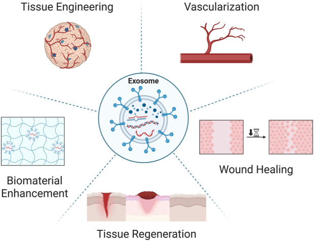
Methods: We present a comprehensive overview of both stem cell-derived and mature-cell-derived exosomes in the context of regenerative vascularization and wound repair, focusing on cargo mechanisms and biomaterial delivery strategies. We also highlight how materials science will be instrumental to both therapeutic delivery and development of fully acellular pro-angiogenic bioengineered exosomes. All cited studies involving exosomes complied with the International Society of Extracellular Vesicles guidelines. To assess the clinical relevance and gaps, we visited clinicaltrials.gov, where keywords "exosome" and "vascular" were searched. Other parameters such as completion status, country, and exosome type further refined our search.

Results: Exosomes were found to promote angiogenesis and improved wound healing outcomes primarily via Vascular Endothelial Growth Factor, FGF2, miR-126, Wnt/β-catenin, Notch and PI3K/Akt pathways. Clinicaltrials.gov revealed only 3 out of 15 completed human exosome studies worldwide related to regenerative vascularization.

Conclusion: Therapies utilizing exosomes as an acellular approach to regenerative vascularization are promising, though challenges with scalability remain. Further mechanistic understanding, standardization, and controlled trials are compulsory prior to widespread human application.

Keywords: angiogenesis; exosomes; regenerative; stem cells; vascularization; wound healing.


Conflict of interest statement

The authors declare that the research was conducted in the absence of any commercial or financial relationships that could be construed as a potential conflict of interest. The author(s) declared that they were an editorial board member of Frontiers, at the time of submission. This had no impact on the peer review process and the final decision.

Figures

FIGURE 1
FIGURE 1
Exosome applications in regenerative medicine. Exosomes, a small class of extracellular vesicles have headlined the regenerative research and medical fields as a novel multifunctional therapeutic. Exosomes have been found to be instrumental in various regenerative hallmarks including tissue engineering, inducible vascularization, accelerated wound healing, tissue regeneration, and enhanced biomaterial efficacy. Created in BioRender. Ravnic, D. (2025) https://BioRender.com/mbrx7wr.
FIGURE 2
FIGURE 2
Exosome biogenesis. The three main classes of extracellular vesicles include: exosomes, microvesicles, and apoptotic bodies. Exosome biogenesis is hallmarked by endosomal invagination resulting in the formation of multi-vesicular bodies (MVBs) which contain intraluminal vesicles (ILVs). The membranes of MVBs eventually fuse with the plasma membrane, which allows for the ILVs to be released from the cell as exosomes. This biogenesis process equips the resulting exosome populations with heterogenous surface markers and encapsulated bioactive cargo. Microvesicles originate from plasma membrane budding, while apoptotic bodies are a result of programmed cell death blebbing. Created in BioRender. Ravnic, D. (2025) https://BioRender.com/mbrx7wr.
FIGURE 3
FIGURE 3
Exosomes and the multicellular wound microenvironment. Proper wound healing is dependent on interdependent multicellular communication within the local microenvironment. (A) The cellular milieu within a healing wound includes local and recruited modulators. These cell types secrete exosomes with diverse cargo including pro-angiogenic and pro-healing cytokines, growth factors, nucleic acids, and adhesion molecules. (B) These exosomes exert significant therapeutic potential in wound repair through facilitating macrophage polarization, microvascular expansion, re-epithelialization, and modulation of the remodeled ECM. (C) An extensive range of studies have demonstrated that exosomes are a useful acellular tool in enhancing the wound healing process via immune cell modulation and increased vascularization. Created in BioRender. Ravnic, D. (2025) https://BioRender.com/mbrx7wr.
FIGURE 4
FIGURE 4
Preclinical applications of exosomes in regenerative medicine. (A) Exosomes have been found to enhance hydrogel efficacy within wound healing through sustained release of pro-angiogenic growth factors and cytokines. (B and C) Direct and indirect methods of exosome engineering have allowed researchers to enhance target specificity while also augmenting the exosomes regenerative capacity through surface alterations or parent cell priming. Created in BioRender. Ravnic, D. (2025) https://BioRender.com/mbrx7wr.

References

    1. Abdik H., Kirbas O. K., Bozkurt B. T., Avsar Abdik E., Hayal T. B., Sahin F., et al. (2024). Endothelial cell-derived extracellular vesicles induce pro-angiogenic responses in mesenchymal stem cells. FEBS Open Bio 14, 740–755. 10.1002/2211-5463.13650 - DOI - PMC - PubMed
    1. Adair, T. H., and Montani J. P. (2010). Angiogenesis. San Rafael (CA): Morgan & Claypool Life Sciences. - PubMed
    1. Alexiadou K., Doupis J. (2012). Management of diabetic foot ulcers. Diabetes Ther. 3, 4. 10.1007/s13300-012-0004-9 - DOI - PMC - PubMed
    1. Antes T. J., Middleton R. C., Luther K. M., Ijichi T., Peck K. A., Liu W. J., et al. (2018). Targeting extracellular vesicles to injured tissue using membrane cloaking and surface display. J. Nanobiotechnol. 16, 61. 10.1186/s12951-018-0388-4 - DOI - PMC - PubMed
    1. Awada H. K., Johnson N. R., Wang Y. (2014). Dual delivery of vascular endothelial growth factor and hepatocyte growth factor coacervate displays strong angiogenic effects. Macromol. Biosci. 14, 679–686. 10.1002/mabi.201300486 - DOI - PMC - PubMed