. 2025 Sep 3;7(5):fcaf326.
doi: 10.1093/braincomms/fcaf326. eCollection 2025.

Biallelic CRELD1 variants cause severe muscle weakness and infantile epilepsy

Affiliations

Biallelic CRELD1 variants cause severe muscle weakness and infantile epilepsy

Manuela D'Alessandro et al. Brain Commun. .

Abstract

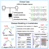
Nicotinic acetylcholine receptors are widely expressed in the peripheral and central nervous systems. Mutations in acetylcholine receptor-subunit genes have been associated with neuromuscular diseases, such as arthrogryposis multiplex congenita (AMC) and epilepsy. We report a patient with arthrogryposis, severe muscle weakness and neurodevelopmental delay. During his first year of life, he developed therapy-refractory epilepsy. Using whole-exome sequencing, we identified the compound pathogenic variants c. 875G>A (p. Cys292Tyr) and c. 959delA (p. Gln320Argfs*25) in the cysteine-rich with epidermal growth factor-like domain protein 1 gene (CRELD1, NM_001077415.3). Recently, functional studies have shown that CRELD1 is a membrane-associated endoplasmic reticulum-resident protein disulphide isomerase that acts as a maturation enhancer of AChR biogenesis, thereby controlling the abundance of functional receptors at the cell surface. To test pathogenicity, we took advantage of the genetics and extremely rapid genome editing in Caenorhabditis elegans. We were able to model these heterozygous variants and observed a decrease in AChRs at the neuromuscular junction. Hence, our study identifies compound heterozygous CRELD1 variants responsible for a rare neurodevelopmental disorder characterized by arthrogryposis, muscle weakness and epilepsy.

Keywords: CRELD1; arthrogryposis; epilepsy; nicotinic acetylcholine receptor.


Conflict of interest statement

The authors report no competing interests.

Figures
Graphical Abstract
Graphical Abstract
Figure 1
Figure 1
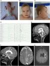
Clinical presentation electroencephalogram and brain MRI. The index patient suffering from severe muscle weakness with limited anti-gravity movements at the age of 8 months (A). Facial stigmata included a prominent forehead, deep-set ears, hypertelorism, broad nose, epicanthus and retrognathia at the age of 14 months (B, C). EEG at 9 months showing the voltage on the Y-axis (each blue bar indicates the scale in microvolts) and the time on the X-axis (the interval between two solid vertical lines corresponds to 1 s). The EEG showed spike-wave complexes singular and in small groups located centroparietal on both sides, but predominantly on the left side. Occasional spread to the temporal (T5) was noted. Generalized spike-slow wave complexes correlated with myoclonic seizures (D). Cranial MRI at 12 months of age showed no major brain malformations (E). Unspecific periventricular hyperintensities (arrows) were noted on T2-weighted (F, G) and on fluid-attenuated inversion recovery (FLAIR) images (H), as well as an enlargement of the outer cerebrospinal fluid space (asterisk; G, H).
Figure 2
Figure 2
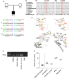
CRELD1 missense variants and expression analysis. Pedigree of the family, illustrating the non-consanguineous parents and the index patient (II/1). Compound heterozygous inheritance was confirmed by Sanger sequencing (A). The amino acid exchange of the missense variant detected in our patient (p. Cys292Tyr) and the missense variant reported in ClinVar (p. Cys192Tyr) affect evolutionarily highly conserved residues. The alignment was performed with Promals3D, consensus_aa = consensus amino acids; more details can be found in Supplementary Methods. (B). Diagram displaying that both cysteines (p. Cys192 and 292) are located in the EGF-like domains and form disulphide bonds that are essential for the domain structure; the CRELD1 EGF-like domains and interchain disulphide bonds were modelled with AlphaFold (https://alphafold.ebi.ac.uk) and visualized with PyMOL. Predicted disulphide bonds are shown in sticks and highlighted in yellow (C). RT-qPCR analysis of total RNA from the patient’s skeletal muscle sample, and from commercial skeletal muscle samples. NTC = no template control. No abnormal splicing was detected on the agarose gel. The full-size uncropped gel is in Supplementary material (D). RT-qPCR results showing downregulation of CRELD1 expression in the patient skeletal muscle compared to the commercial skeletal muscle as well as the variable CRELD1 expression in the brain, fetal brain and skeletal muscle tissues (E). Each data point represents the average of three technical replicates. For the patient sample, N = 4; for the other samples, N = 3. Sk. = skeletal.
Figure 3
Figure 3
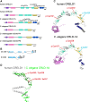
Domain architecture of the human, C. elegans and D. melanogaster CRELD proteins. Human CRELD1 (NP_001070883.2) consists of an N-terminal signal peptide (violet), the WE domain (pink) specific for the CRELD protein family, two EGF-like domains (green) followed by an EGF-like-cbEGF tandem domain consisting of an EGF-like and a Ca2+-binding EGF-like (cbEGF) domain (yellow) and two C-terminal transmembrane domains (blue) (A). The human CRELD2 gene encodes a homologous protein lacking only the transmembrane domains (A). C. elegans and D. melanogaster have only one CRELD gene, but express two different alternatively spliced isoforms, one with and one without the transmembrane domains (A, B). The alternative splicing of the last exons in C. elegans (exon 9a or 9b) and Drosophila (exon PA or PB) gives rise to the two different CRELD1 isoforms (B). The domain architecture is predicted to be similar to human CRELD1, which contains an additional EGF-like domain (A). Modelling of the protein structure with AlphaFold and visualization with Pymol of human CRELD1 and C. elegans CRLD-1A highlight the similar tertiary structure. Cysteines and disulphide bonds are shown in sticks. The cysteine mutated in our patient forms a disulphide bond in the third EGF-like domain. Human Cys 192 and Cys 292 and C. elegans Cys 187 and Cys 228 are highlighted in red. Human Gln 320 and C. elegans Ile 257 are highlighted in cyan (C). AlphaFold predicted structures alignment using ChimeraX16.1. The C-terminal domains encompassing the cbEGF and the two transmembrane domains from both human CRELD1 and C. elegans CRLD-1A were aligned. Human CRELD1 (from Asp 247 to Arg 420 residue) is shown in grey, C. elegans CRLD-1A (from Ile 192 to Tyr 356) is in green. Human Cys 292 and Gln 320 and C. elegans Cys 228 and Ile 257 are highlighted in red (D).
Figure 4
Figure 4
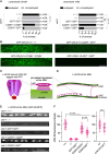
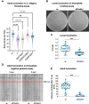
The patient’s compound heterozygous variant affects L-AChR expression at the NMJ. Levamisole test on both wild types (indicated as +/+), crld-1 knock outs (indicated as −/−), crld-1 Cys228Tyr and Ile257RFs*25 knock-ins (indicated as C228Y/C228Y and I257*/I257*, respectively) and crld-1 compound heterozygous mutants (indicated as C228Y/I257*). C228Y/I257* mutants are largely resistant to levamisole. Experiments were repeated six times. ns = not significant P = 0.4979 for C228Y/C228Y on 1 mM levamisole, ****P < 0.0001, after Bonferroni correction, Fisher’s exact probability test. Grey bars indicate the percentage of moving animals after overnight exposure to either 1 mM or 0.6 mM levamisole, and black bars indicate the percentage of paralyzed animals, the number of animals tested for each genotype is indicated (A). Distribution of GFP-CRLD-1A in muscle cells of gfp-crld-1a knock-in animals carrying also the different human CRELD1 mutations found in the patient. In compound heterozygous mutants (C228Y/I257*), the GFP-CRLD-1a pattern is much less intense as compared to the control (+/+). Scale bars = 10 μm (B). Schematic representation of L-AChR in which the subunit UNC-29 is tagged with RFP (C). Organization of excitatory cholinergic NMJs in C. elegans. Muscle cells (green) are innervated by cholinergic motoneurons (purple). L-AChR are clustered in front of cholinergic boutons (inset) along the VNC and DNC. (D). Confocal imaging of the L-AChR reporter UNC-29::tagRFP at the ventral nerve cords of control (+/+), crld-1(I257*) and crld-1(C228Y) alone or crossed with crld-1(I257*) mutant worms (C228Y/I257*). Scale bars = 10 μm (E). Quantification of UNC-29::tagRFP fluorescence at the ventral nerve cords. Each dot represents the fluorescence intensity of UNC-29::tagRFP measured at the VNC of a worm. Data are normalized on control mean and presented as mean ± SD; control, +/+: n = 43 animals, crld-1(−/−): n = 37 animals, crld-1(I257*/I257*): n = 40 animals, crld-1(C228Y/C228Y): n = 57 animals, crld-1(C228Y) crossed with crld-1(I257*), C228Y/I257*: n = 41; experiments were repeated four times. ****P < 0.0001, Kruskal–Wallis test. Mann–Whitney test was applied to compare C187Y/C187Y animals (n = 35 animals) with their control (n = 48 animals). ***P < 0.001 (F).
Figure 5
Figure 5
CRELD is required for locomotion in both C. elegans and Drosophila models. Thrashing assay for C. elegans locomotion: body bends frequency at day 1 of adulthood of wild types (indicated as +/+), crld-1 knockouts (indicated as −/−), crld-1 Cys228Tyr and Ile257RFs*25 knock-ins (indicated as C228Y/C228Y and I257*/I257*, respectively) and crld-1 compound heterozygous mutants (indicated as C228Y/I257*). Black line: median value, dotted lines: lower and upper quartiles, n: number of animals scored in at least three independent experiments; ns = not significant, ****P < 0.0001, Kruskal–Wallis test (A). Crawling assay for Drosophila larval locomotion: single larvae are semi-automatically tracked (green line represents distance crawled in 20 s) (B). Speed in mm/s. Dots represent individual larvae, w-n = 39, dCreld−/− n = 37 (C). Negative geotaxis assay for adult locomotion: flies are placed in glass tubes and tapped on the ground. Climbing is recorded (D). Distance from the fly head to the bottom after 6 s. Boxes in box plots represent the interquartile range and median; whiskers represent the minimum and maximum. Significance tested by unpaired, two-tailed Student’s t-test assuming heteroscedasticity (*** indicates P < 0.001). Dots represent individual animals, w-n = 45, dCreld−/− n = 45 (E). In panels C and E, each experiment was performed in at least three independent biological replicates.

References

    1. Albuquerque EX, Pereira EF, Alkondon M, Rogers SW. Mammalian nicotinic acetylcholine receptors: From structure to function. Physiol Rev. 2009;89(1):73–120. - PMC - PubMed
    1. Colombo SF, Mazzo F, Pistillo F, Gotti C. Biogenesis, trafficking and up-regulation of nicotinic ACh receptors. Biochem Pharmacol. 2013;86(8):1063–1073. - PubMed
    1. Hurst R, Rollema H, Bertrand D. Nicotinic acetylcholine receptors: From basic science to therapeutics. Pharmacol Ther. 2013;137(1):22–54. - PubMed
    1. Zoli M, Pucci S, Vilella A, Gotti C. Neuronal and extraneuronal nicotinic acetylcholine receptors. Curr Neuropharmacol. 2018;16(4):338–349. - PMC - PubMed
    1. Gotti C, Clementi F. Neuronal nicotinic receptors: From structure to pathology. Prog Neurobiol. 2004;74(6):363–396. - PubMed

LinkOut - more resources