. 2025 Jul 8;10(9):3044-3057.
doi: 10.1016/j.ekir.2025.06.056. eCollection 2025 Sep.

A New Prognostic Score Based on Cell-Mediated Immunity for Cytomegalovirus Infection After Transplantation

Affiliations

A New Prognostic Score Based on Cell-Mediated Immunity for Cytomegalovirus Infection After Transplantation

Delphine Kervella et al. Kidney Int Rep. .

Abstract

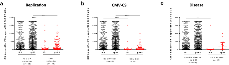
Introduction: The interferon gamma (IFN-γ) enzyme-linked immunosorbent spot is a highly sensitive immune assay that enables the assessment of cytomegalovirus (CMV)-specific cell-mediated immunity (CMI) and can identify at-risk transplant patients of CMV infection; however, its clinical implementation remains elusive.

Methods: We developed a novel CMV-CMI risk-score based on the standardized T-SPOT.CMV assay against 2 CMV antigens (immediate-early protein 1 [IE-1] and 65 kDa phosphoprotein [pp65]), a biomarker predicting CMV infection, both high viral replication, and disease by performing a pooled analysis of 570 kidney transplants participating in different clinical trials and subsequently validating it in 146 consecutives solid organ transplants (SOT) in an interventional trial. By incorporating clinical variables into the CMV-CMI risk-score, we built an integrative prognostic system quantifying the risk of CMV infection (CMV-PrognosTIC score) using elastic net penalized regression analysis.

Results: In the pooled derivation cohort, whereas specific IE-1/pp65-specific CMV-CMI frequencies independently correlated with high risk of CMV infection (areas under the curve [AUCs]: 0.694, P < 0.0001; 0.719, P < 0.0001, respectively), by combining both responses, 3 CMV-CMI risk-scores appeared, accurately discriminating low-risk (LR) from intermediate-risk (IR) and high-risk (HR) patients (98.7% negative predictive value [NPV], 97.2% sensitivity). Its prospective implementation guiding decision-making in an independent SOT cohort confirmed the very high NPV and sensitivity identifying LR patients. By integrating type of preventive therapy, patient age, and donor (D) and recipient (R) CMV-serostatus to the CMV-CMI risk-score, we generated a global risk-prognostic model showing accurate discrimination and calibration in both derivation (AUC: 0.807) and validation cohorts (AUC: 0.719).

Conclusion: We developed a robust CMV-PrognosTIC score to quantify the risk of CMV infection in SOT, which may be readily implemented in clinical transplantation to personalize CMV preventive therapies.

Keywords: CMV infection; cell-mediated immunity; monitoring; solid organ transplantation.


Figures

None
Graphical abstract
Figure 1
Figure 1
Flow-chart of the study. D+, CMV IgG seropositive donor; HR, high-risk CMI; IR, intermediate-risk CMI; LR, low-risk CMI; R−, CMV IgG seronegative recipient; R+, CMV IgG seropositive-recipient; SOT, solid organ transplants. ∗Patients excluded from validation cohort because of loss of follow-up (n = 13), use of antirejection therapy (n = 7), unavailable blood samples (n = 7), and prophylaxis initiation before the CMV-CMI test (n = 6).
Figure 2
Figure 2
CMV-CMI results in the derivation cohort. CMV-CMI for patients developing (a) CMV replication, (b) CMV-CSI, and (c) CMV disease. (a) Any CMV replication versus no replication among all patients: 2.0 (0.0–16.0) versus 10.0 (0.0–118.0) median IE-1-specific IFN-γ spots/2.5 × 105 PBMC respectively, P < 0.0001, and 27.0 (1.0–123.0) vs. 109.0 (4.0–332.0) median pp65-specific IFN-γ spots/2.5 × 105 PBMC, respectively; P < 0.0001. (b) CMV-CSI versus no CMV-CSI among all patients: 1.0 (0.0–4.0) versus 10.0 (0.0–99.0) median IE-1-specific IFN-γ spots/2.5 × 105 PBMC respectively; P < 0.0001, and 6.0 (0.0–55.0) versus 109.0 (6.0–323.0) versus median pp65-specific IFN-γ spots/2.5 × 105 PBMC respectively; P < 0.0001. (c) CMV disease versus no CMV disease among all patients: 2.5 (0.0–13.3) versus 10.0 (0.0–99.0) median IE-1-specific IFN-γ spots/2.5 × 105 PBMC, respectively; P = 0.0424, and 68.2 (3.8–175.8.0) versus 109.0 (6.0–323.0) median pp65-specific IFN-γ spots/2.5 × 105 PBMC respectively, P = 0.2903). CMI, cell-mediated immunity; CMV, cytomegalovirus; CSI, clinically significant infection; D+, CMV IgG seropositive donor; IE-1, immediate-early protein 1; IFN-γ, interferon gamma; PBMC, peripheral blood mononuclear cell; pp65, 65 kDa phosphoprotein; R–, CMV IgG seronegative recipient; R+, CMV IgG seropositive-recipient.
Figure 3
Figure 3
Risk-score classification with IE-1 and pp65-specific CMV-CMI. (a) ROC curves for the risk of CMV replication. (b) ROC curves for the risk of CMV-CSI. (c) Classification of patients in 3 distinct risk categories considering the combination of both IE-1 and pp65-specific CMI (CMV-CMI risk-score): low-risk (LR), intermediate-risk (IR), and high-risk (HR). CMI, cell-mediated immunity; CMV, cytomegalovirus; CSI, clinically significant infection; IE-1, immediate-early protein 1; pp65, 65 kDa phosphoprotein; ROC, receiver operating characteristics.
Figure 4
Figure 4
CMV infection rates according to CMV-CMI risk stratification in the derivation cohort. Kaplan-Meier CMV infection-free survival curves for (a, b, c) any CMV replication, (c, d, e) CMV-CSI, and (g, h, i) CMV disease in all patients of the derivation cohort according to (a, d, g) IE-1-specific CMV-CMI, (c, f, i) pp65-specific CMV-CMI and for the combined IE-1 and pp65-specific CMV-CMI risk-score. CMI, cell-mediated immunity; CMV, cytomegalovirus; CSI, clinically significant infection; IE-1, immediate-early protein 1; pp65, 65 kDa phosphoprotein.
Figure 5
Figure 5
Kaplan-Meier CMV CSI-free survival curves in the validation cohort. (a) KM CMV-CSI-free survival curves between different CMI-CMV risk-scores and (b) when stratified according to the prevention strategies used. CMI, cell-mediated immunity; CMV, cytomegalovirus; CSI, clinically significant infection; HR, high-risk CMI; IR, intermediate-risk CMI; LR, low-risk CMI.
Figure 6
Figure 6
ROC curves of each variable and combination of variables for the risk of CMV-CSI, with respective AUC. AUC, area under the curve; CMV-CSI, cytomegalovirus clinically significant infection; ROC, receiver operating characteristics. ROC curves were compared using the DeLong method.
Figure 7
Figure 7
CMV-PrognosTIC score performances. ROC curves of the CMV-PrognosTIC score in the (a) derivation and (b) validation cohorts. (c) Calibration plot of the CMV-PrognosTIC score for the validation cohort. The graph represents the ratio of positive cases and the standard error of the observed risk of CMV-CSI (Y axis) according to each centile of CMV-CSI risk predicted by the CMV-CMI PrognosTIC score (x axis), illustrating the fit between expected and observed risk. Perfect calibration is represented by the line. We observe good predictions along all the risk percentiles, with certain overdiagnosis of high-risk CMV-CMI for CSI (patient with a lower observed risk than the predicted risk when the predicted risk is high). Density plot of the patients from the (d) derivation cohort and (e) validation cohort. Each model value (x axis) is associated with a risk of CMV-CSI (dash lines representing different values of CMV-CSI risk). Density of patients for each model value is represented on the graph, stratifying patients in 3 categories according to their CMV-CMI risk-score: LR (green), IR (blue) and HR (red). Furthermore, individual patient’s model value given by the CMV-Prognostic score is represented on the x-axis, characterizing each patient by the occurrence (red) or not (black) of a CMV-CSI during follow-up. CMI, cell-mediated immunity; CMV, cytomegalovirus; CSI, clinically significant infection; HR, high-risk CMI; IE-1, immediate-early protein 1; IR, intermediate-risk CMI; LR, low-risk CMI; pp65, 65 kDa phosphoprotein.

References

    1. Raval A.D., Kistler K.D., Tang Y., Murata Y., Snydman D.R. Epidemiology, risk factors, and outcomes associated with cytomegalovirus in adult kidney transplant recipients: a systematic literature review of real-world evidence. Transpl Infect Dis. 2021;23 doi: 10.1111/tid.13483. - DOI - PubMed
    1. Kotton C.N., Kumar D., Caliendo A.M., et al. The third international consensus guidelines on the management of cytomegalovirus in solid-organ transplantation. Transplantation. 2018;102:900–931. doi: 10.1097/TP.0000000000002191. - DOI - PubMed
    1. Bestard O., Kaminski H., Couzi L., Fernández-Ruiz M., Manuel O. Cytomegalovirus cell-mediated immunity: ready for routine use? Transpl Int. 2023;36 doi: 10.3389/ti.2023.11963. - DOI - PMC - PubMed
    1. Lúcia M., Crespo E., Cruzado J.M., Grinyó J.M., Bestard O. Human CMV-specific T-cell responses in kidney transplantation; toward changing current risk-stratification paradigm. Transpl Int. 2014;27:643–656. doi: 10.1111/tri.12318. - DOI - PubMed
    1. Griffiths P., Reeves M. Pathogenesis of human cytomegalovirus in the immunocompromised host. Nat Rev Microbiol. 2021;19:759–773. doi: 10.1038/s41579-021-00582-z. - DOI - PMC - PubMed