

Use of dual-energy X-ray absorptiometry to evaluate variation in bone shape and alignment associated with radiographic knee osteoarthritis: Findings from a study of 19,053 individuals in UK Biobank
- PMID: 40980743
- PMCID: PMC12445559
- DOI: 10.1016/j.ocarto.2025.100667
Use of dual-energy X-ray absorptiometry to evaluate variation in bone shape and alignment associated with radiographic knee osteoarthritis: Findings from a study of 19,053 individuals in UK Biobank
Abstract
Objective: Lower limb alignment may predispose to, or exacerbate, symptoms of knee osteoarthritis. To examine the role of this and other joint shape variation, we conducted a cross-sectional study investigating relationships between radiographic knee osteoarthritis (rKOA) and knee shape in dual-energy X-ray absorptiometry (DXA) images from UK Biobank (UKB).
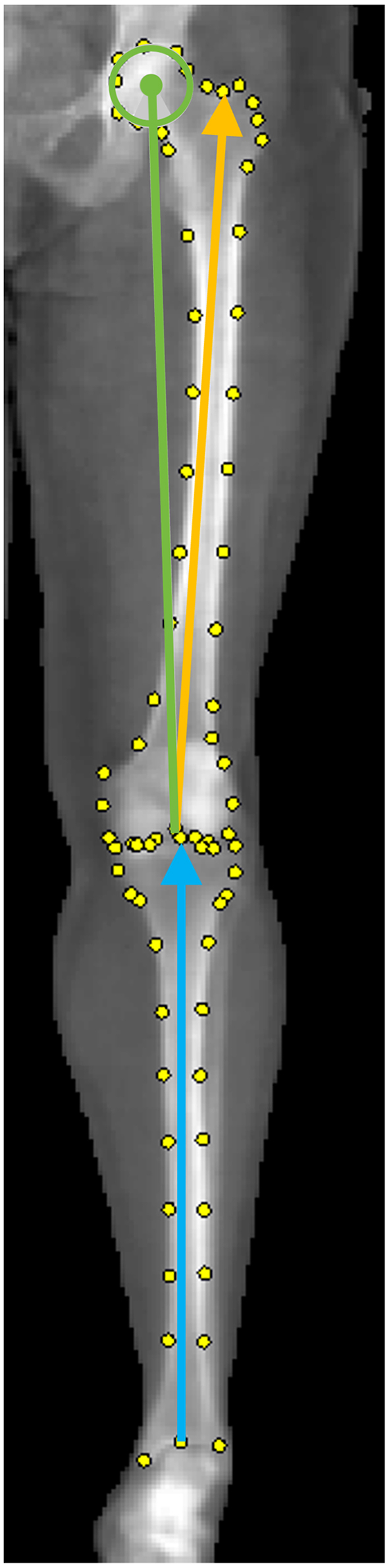
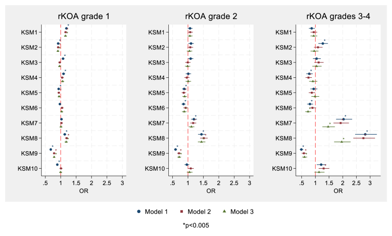
Methods: Associations between the first ten knee shape modes (KSMs), derived from statistical shape modelling, and rKOA grade were analysed using logistic regression, adjusting for age, sex, height, weight, and ethnicity. An additional model included adjustment for hip-knee-ankle (HKA) angle, derived from total body DXA scans, to reflect knee alignment. Composite figures illustrate knee shape characteristics associated with each rKOA grade.
Results: 19,053 individuals were included (mean 63.7 years, 48 % males), of whom 80.7 %, 14.6 %, 3.6 % and 1.2 % had rKOA grades 0, 1, 2 and 3-4, respectively. Several KSMs were associated with rKOA in confounder-adjusted analyses, with higher grades showing stronger relationships. These associations were attenuated by adjustment for HKA. As expected, composite shape models revealed that higher rKOA grades were associated with greater varus malalignment. After HKA adjustment, composite shape models showed less varus alignment, with other shape differences, such as altered proximal tibial metaphysis and lateral patella displacement, emerging in higher-grade rKOA.
Conclusions: Our cross-sectional analyses between joint shape and DXA-derived rKOA grade showed expected relationships with varus malalignment, which were attenuated after adjusting for HKA. Other shape differences, particularly in higher-grade rKOA, emerged independently of alignment, warranting further investigation.
Keywords: Dual-energy X-ray absorptiometry; Knee alignment; Knee osteoarthritis; Statistical shape modelling.
© 2025 The Author(s).
Conflict of interest statement
No competing financial interests exist.
Figures


References
-
- Sharma L., Song J., Felson D.T., Cahue S., Shamiyeh E., Dunlop D.D. The role of knee alignment in disease progression and functional decline in knee osteoarthritis. JAMA. 2001;286(2):188–195. - PubMed
-
- Brouwer G.M., van Tol A.W., Bergink A.P., Belo J.N., Bernsen R.M., Reijman M., et al. Association between valgus and varus alignment and the development and progression of radiographic osteoarthritis of the knee. Arthritis Rheum. 2007;56(4):1204–1211. - PubMed
LinkOut - more resources
Full Text Sources
Research Materials
