. 2025 Sep 23;10(1):307.
doi: 10.1038/s41392-025-02391-9.

Ninjurin-1 mediates cell lysis and detrimental inflammation of PANoptosis during influenza A virus infection

Affiliations

Ninjurin-1 mediates cell lysis and detrimental inflammation of PANoptosis during influenza A virus infection

Yitian Xu et al. Signal Transduct Target Ther. .

Abstract

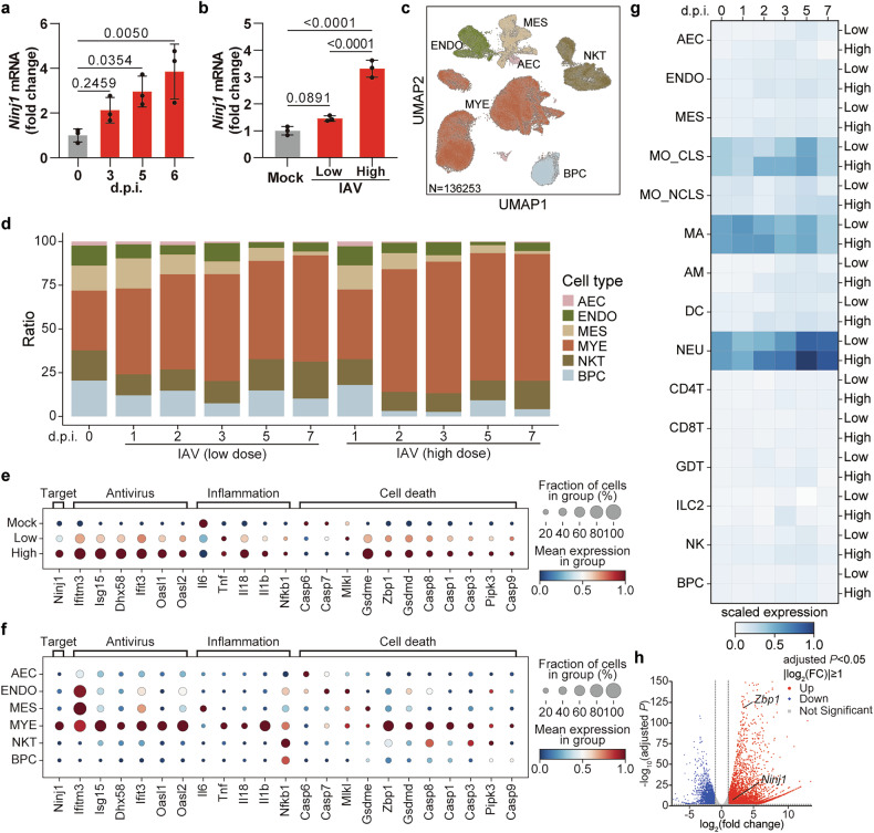
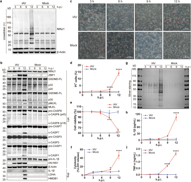
Influenza A virus (IAV) induces ZBP1-mediated PANoptosis, a form of lytic inflammatory cell death characterized by concurrent activation of the pyroptosis, necroptosis and apoptosis pathways. Ninjurin-1 (NINJ1) is a recently identified mediator of plasma membrane rupture but functions diversely in different types of cell death. However, little is known about the role of NINJ1 in IAV-induced PANoptosis and viral pneumonia. Here, we report that IAV infection triggered an increase in the expression of NINJ1, which then oligomerized and mediated cell lysis in infected macrophages. The deficiency of NINJ1 prevented plasma membrane rupture and the release of DAMPs and IL-1β without affecting the progression of cell death. Activation of any single PANoptosis pathway was sufficient to trigger the oligomerization of NINJ1 and robust cell lysis. Accordingly, only when all PANoptosis pathways were concurrently blocked could the oligomerization of NINJ1, cell death, and cell rupture be prevented. Ablation of NINJ1 in vivo also alleviated IAV-induced lung injury and mortality. Furthermore, we revealed an association between NINJ1 upregulation and poor outcomes in patients with COVID-19. Collectively, our findings indicate a pivotal role of NINJ1 in the immunopathology of IAV infection and its potential as a bioindicator of disease severity and prognosis in viral pneumonia and viral sepsis.


Conflict of interest statement

Competing interests: The authors declare no competing interests.

Figures

Fig. 1
Fig. 1
NINJ1 is upregulated upon IAV infection. a, b qRT‒PCR analysis of relative Ninj1 expression in murine lungs at indicated dpi (1×10⁵ PFU) (a) or at 5 dpi infected with low (1×10³ PFU) or high (1×10⁵ PFU) IAV dose (b). c UMAP of 136,253 single cells from lungs at indicated dpi with low/high IAV dose. AEC, alveolar epithelial cell; ENDO, endothelial cell; MES, mesenchymal cell; MYE, myeloid cell; NKT, NK cell and T cell; BPC, B cell and plasma cell. d Proportion of major cell clusters by group and dpi. e, f Dot plots indicating the relative expression of indicated genes in different groups (e) or in different cell clusters (f). g Matrix plot of Ninj1 expression per cell cluster at different dpi with low/high IAV dose. MO_CLS, classical monocyte; MO_NCLS, non-classical monocyte; MA, macrophage; AM, alveolar macrophage; DC, dendritic cell; NEU, neutrophil; CD4T, CD4+ T cell; CD8T, CD8+ T cell; GDT, γδ T cell; ILC2, group 2 innate lymphoid cell; NK, natural killer cell. h Volcano plot of 1,028 differentially expressed genes in IAV- and mock-infected BMDMs at 12 hpi. Red and blue dots represent 4847 upregulated and 2813 down-regulated genes respectively. n = 3 biological replicates/group. Data are representative of at least two independent experiments (a, b) and are presented as mean ± SD. Analysis was performed via one-way ANOVA (a, b)
Fig. 2
Fig. 2
Oligomerization of NINJ1 synchronizes with PANoptosis during IAV infection. a, b Immunoblots of indicated proteins in BMDMs at indicated h.p.i. (hours post infection), BS3-crosslinked (a) or non-crosslinked (b). FL, full length; c-, cleaved; Lys., lysates; Sup., supernatants. cf Representative images (c) and quantification (d) of PI+ cells, cell viability (e), and LDH release (f) in BMDMs treated as panel (a, b). Scale bars, 100 μm. g Silver staining of supernatants from (b). h, i Concentrations of IL-1β (h) and TNF-α (i) in supernatants from (f). Data are representative of three independent experiments and are presented as mean ± SD. Two-way ANOVA was used. *p < 0.05; ****p < 0.0001
Fig. 3
Fig. 3
NINJ1 mediates IAV-induced cell lysis without compromising cell death. a, b Representative images (a) and quantification (b) of PI+ cells in wild-type (WT) and Ninj1-/- BMDMs at 12 hpi. The arrows indicate unlysed cells exhibiting a balloon-like morphology. Scale bars, 100 μm and 40 μm in the enlarged image. cf LDH release (c), immunoblots of the indicated proteins (d), silver staining of supernatants (e), and cell viability (f) in BMDMs (genotypes as above) at 12 hpi. g Concentrations of TNF-α in supernatants from WT and Ninj1-/- BMDMs at 16 hpi. h Concentrations of IL-1β in supernatants from WT, Ninj1-/-, and Gsdmd-/- BMDMs at 16 hpi. i qRT‒PCR analysis of IAV-NP vRNA in WT and Ninj1-/- BMDMs at 12 hpi (normalized to WT group). j Immunoblots of NINJ1 in BMDMs with/without glycine (10 mM) at 12 hpi, BS3-crosslinked. k qRT‒PCR analysis of Ninj1 expression in BMDMs with/without glycine (10 mM) at 12 hpi (normalized to uninfected controls). l LDH release in WT and Ninj1-/- BMDMs with/without glycine (10 mM) at 12 hpi. m Cell viability of BMDMs with/without glycine (10 mM) at 12 hpi. Data are representative of at least two independent experiments and are presented as mean ± SD. Student’s t-test (f, i, and m) or two-way ANOVA (b, c, g, h, k, and l) was applied. ns, not significant; ****p < 0.0001
Fig. 4
Fig. 4
NINJ1-mediated cell lysis depends on ZBP1. a, b Representative images (a) and quantification (b) of PI+ cells of WT, Zbp1-/- and Ifnar1-/- BMDMs at 16 hpi. Scale bars, 100 μm. c, g Immunoblots of the indicated proteins in the BMDMs (genotypes as above) at 16 hpi, BS3-crosslinked (g) or non-crosslinked (c). d Cell viability of WT, Ninj1-/-, Zbp1-/- and Ifnar1-/- BMDMs at 16 hpi. e LDH release in WT, Zbp1-/-, and Ifnar1-/- BMDMs with/without glycine (10 mM) at 16 hpi. f Silver staining of supernatants from (c). h, i LDH (h) and IL-1β (i) release in WT, Ninj1-/-, and Zbp1-/- BMDMs at 16 hpi. Data are representative of three independent experiments and are presented as mean ± SD. Kruskal‒Wallis test (d) or two-way ANOVA (b, e, h, and i) was applied. ns, not significant; **p < 0.01; ***p < 0.001; and ****p < 0.0001
Fig. 5
Fig. 5
Activation of any PANoptosis pathway induces NINJ1 oligomerization during IAV infection. a, d Immunoblots of NINJ1 in BMDMs of indicated genotypes at 16 hpi, BS3-crosslinked. b Viability of BMDMs of indicated genotypes at 16 hpi. c LDH release in BMDMs of indicated genotypes at 16 hpi, with/without glycine (10 mM). e, g Immunoblots of indicated proteins in WT, Gsdmd-/-Gsdme-/-, and Gsdmd-/-Gsdme-/-Mlkl-/- BMDMs at 16 hpi, treated with DMSO, Z-IETD-FMK (25 μM), or Z-VAD-FMK (25 μM), BS3-crosslinked (g) or non-crosslinked (e). f Representative images of PI+ cells from panel (e). Scale bars, 100 μm. See also Supplementary Fig. 5f. h, i Cell viability (h) and LDH release (i) in WT, Gsdmd-/-Gsdme-/-, and Gsdmd-/-Gsdme-/-Mlkl-/- BMDMs at 16 hpi, treated with DMSO, Z-IETD-FMK, Z-VAD-FMK, or Z-VAD-FMK plus glycine. Data are representative of three independent experiments and are presented as mean ± SD. Kruskal‒Wallis test (b) or two-way ANOVA (c, h, and i) was applied. ns, not significant; **p < 0.01; and ****p < 0.0001
Fig. 6
Fig. 6
NINJ1 drives IAV-induced lung pathology and hyperinflammation. ac Survival curves of (a) Ninj1-/- mice and their littermates; (b) Zbp1-/- and WT mice; (c) Gsdmd-/-, Gsdme-/-, Mlkl-/-, and WT mice. d, e H&E staining (d) and pathological scores (e) of the same lung lobe of WT and Ninj1-/- mice at 5 dpi. The right panel in (d) shows magnified images of the insects. Scale bars, 2 mm and 0.2 mm (insects). f Viral titers in lungs of WT, Ninj1-/- and Zbp1-/- mice at 5 dpi. g Cellular pellets in BALF of WT and Ninj1-/- mice at 8 dpi after centrifugation. hl, o Concentrations of total proteins (h) and indicated cytokines (il, o) in BALF of WT and Ninj1-/- mice at 3, 6, and 8 dpi. m, n Frequencies of CD45+ cells (m) and Ly6C+ monocytes (n) among total live cells in BALF of WT and Ninj1-/- mice at 3 and 6 dpi. Gating strategies are shown in Supplementary Fig. 9a. Mice were infected with LD100 (a, c) or LD50 (b, do) of IAV. Data are representative of at least two independent experiments and are presented as mean ± SD. Two-way ANOVA (ho), one-way ANOVA (f), or Student’s t-test (e) was used. Survival curves were analyzed by the log-rank test (ac)

References

    1. Cline, T. D., Karlsson, E. A., Seufzer, B. J. & Schultz-Cherry, S. The hemagglutinin protein of highly pathogenic H5N1 influenza viruses overcomes an early block in the replication cycle to promote productive replication in macrophages. J. Virol.87, 1411–1419 (2013). - PMC - PubMed
    1. Marvin, S. A., Russier, M., Huerta, C. T., Russell, C. J. & Schultz-Cherry, S. Influenza virus overcomes cellular blocks to productively replicate, impacting macrophage function. J. Virol.91, e01417–16 (2017). - PMC - PubMed
    1. Malireddi, R. K. S., Kesavardhana, S. & Kanneganti, T. D. ZBP1 and TAK1: master regulators of NLRP3 inflammasome/pyroptosis, apoptosis, and necroptosis (PAN-optosis). Front. Cell Infect. Microbiol.9, 406 (2019). - PMC - PubMed
    1. Zheng, M. & Kanneganti, T. D. The regulation of the ZBP1-NLRP3 inflammasome and its implications in pyroptosis, apoptosis, and necroptosis (PANoptosis). Immunol. Rev.297, 26–38 (2020). - PMC - PubMed
    1. Zheng, M., Karki, R., Vogel, P. & Kanneganti, T. D. Caspase-6 is a key regulator of innate immunity, inflammasome activation, and host defense. Cell181, 674–687.e613 (2020). - PMC - PubMed

MeSH terms

LinkOut - more resources