. 2025 Sep 23;11(1):76.
doi: 10.1038/s41421-025-00825-w.

MT1-MMP inhibition rejuvenates ageing brain and rescues cognitive deficits in obesity

Affiliations

MT1-MMP inhibition rejuvenates ageing brain and rescues cognitive deficits in obesity

Pallavi Asthana et al. Cell Discov. .

Abstract

Obesity has been linked to an increased risk of cognitive impairment and dementia in later life. Although aging and obesity are both associated with cognitive decline, it remains unclear how they interact to affect cognitive function across the lifespan and how brain function might mediate their relationship with cognition. Our previous findings and other studies have shown that membrane type 1-matrix metalloproteinase (MT1-MMP/MMP14), which increases with age, regulates energy homeostasis. Inhibiting MT1-MMP improves insulin sensitivity, reduces body fat, and lowers serum cholesterol. Here, we demonstrate that MT1-MMP links neuroinflammation to cognitive decline in aging and obesity. Inflammatory responses in the brain increase MT1-MMP activation in the hippocampus of both mice and humans. Activation of hippocampal MT1-MMP alone can trigger cognitive decline and synaptic impairment independently of neuroinflammation. Conversely, ablation of MT1-MMP in the hippocampus reverses cognitive decline and improves synaptic plasticity in aging and obesity. Pharmacological inhibition of MT1-MMP, through an orally administered brain-penetrant inhibitor or targeted delivery of a neutralizing antibody to the hippocampus, improves memory and learning in aged and obese mice without toxicity. Mechanistically, MT1-MMP proteolytically inactivates G-protein-coupled receptor 158 (GPR158), a hippocampal receptor for osteocalcin (OCN) that is important for the maintenance of cognitive integrity, thus suppressing the ability of the OCN-GPR158 axis to promote cognition in aging and obesity. These findings suggest a new mechanism underlying hippocampal dysfunction and reveal the potential for treating multiple age-related diseases, including neurodegenerative disorders, obesity, diabetes, and atherosclerosis, with a single MT1-MMP-blocking agent.


Conflict of interest statement

Conflict of interest: The authors declare no competing interests.

Figures

Fig. 1
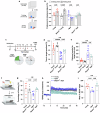
Fig. 1. Age-related neuroinflammation drives MT1-MMP activation in the hippocampus.
a Immunoblotting of MT1-MMP from hippocampal lysates of 2-month-old (young) and 18-month-old (aged) mice (n = 2 mice per group). b Quantification of MT1-MMP expression relative to β-actin loading control (n = 5 mice per group). Two-tailed unpaired Student’s t-test. c MMP14 activity was measured as relative fluorescence units (RFU) in the hippocampus sample of young and aged mice. Two-tailed unpaired Student’s t-test (n = 5 mice per group). d MMP14 normalized transcript per million (nTPM) data from adult genotype tissue expression (GTEx) portal and comparative analysis between young and aged human hippocampus tissue. Two-tailed unpaired Student’s t-test. e Hippocampal Mmp14 mRNA levels by qPCR in young, aged, and C52-treated aged mice. One-way ANOVA followed by Tukey’s multiple comparisons test. (n = 5 young, n = 6 aged, n = 6 aged + C52 mice). fm Correlation analysis between MMP14 and pro-inflammatory factors NF-κB (f), IL-1B (g), IL-6 (h), CCL3 (i), IL23A (j), CD11B (k), TNF (l) and C1QB (m) from the RNA sequencing data from human hippocampus tissues obtained through GTEx portal. Data are expressed as mean ± SEM, and each data point represents individual mouse/human.
Fig. 2
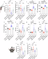
Fig. 2. Systemic MT1-MMP depletion alleviates aging-associated cognitive impairment.
a Schematic diagram of object location test. b Time spent exploring the displaced object expressed as the preference for the displaced object (%) from 2-month-old (young) to 18-month-old (aged) WT and Mmp14+/− mice. One-way ANOVA followed by Fisher’s LSD post hoc test (n = 6 mice per group). c Schematic diagram of Barnes maze test. d, e Percentage of total time spent in the target quadrant (d) and distance traveled to the target hole (e) in Barnes maze test. One-way ANOVA followed by Tukey’s multiple comparisons test (n = 10 young, n = 10 Mmp14+/− young, n = 11 aged, n = 7 Mmp14+/− aged mice). f Schematic diagram of contextual fear conditioning test. g Percentage of freezing time during a contextual fear conditioning test performed by young and aged WT and Mmp14+/− mice. One-way ANOVA followed by Fisher’s LSD post hoc test (n = 6 mice per group). h, i LTP in the CA1 hippocampal region quantified as the change in fEPSP (h) and the average of fEPSP (i) from young and aged WT and Mmp14+/− mice. One-way ANOVA followed by Tukey’s multiple comparisons test (n = 5 mice per group). Data are expressed as mean ± SEM, and each data point represents individual mouse.
Fig. 3
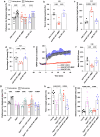
Fig. 3. Selective ablation of MT1-MMP in the hippocampus reverses cognitive impairment.
a Preference for the displaced object in object location task by 2-month-old (young) and 18-month-old (aged) mice injected with shRNA targeting Mmp14 (shMMP14) or scrambled shRNA (shControl) in the DG region of the hippocampus. One-way ANOVA followed by Tukey’s multiple comparisons test (n = 7 young + shControl, n = 7 young + shMMP14, n = 6 aged + shControl, n = 6 aged + shMMP14). b, c Percentage of total time spent in the target quadrant (b) and distance traveled to the target hole (c) in the Barnes maze test. One-way ANOVA followed by Tukey’s multiple comparisons test (n = 8 mice per group). d Percentage of freezing time during a contextual fear conditioning test. One-way ANOVA followed by Fisher’s LSD post hoc test (n = 6 mice per group). e, f Change in fEPSP in the hippocampal CA1 for LTP analysis (e) and the average of fEPSP (f). One-way ANOVA followed by Tukey’s multiple comparisons test (n = 5 young + shControl, n = 5 young + shMMP14, n = 6 aged + shControl, n = 5 aged + shMMP14). Data are expressed as mean ± SEM, and each data point represents individual mouse.
Fig. 4
Fig. 4. Pharmacological inhibition of MT1-MMP improves memory function and metabolism in 18-month-old aged mice.
a, b Cognitive function assessment using the Y-maze task (a) and contextual memory assessment (b) by examining the percentage of time freezing. One-way ANOVA followed by Tukey’s multiple comparisons test (n = 8 young + vehicle, n = 8 young + Ro 28-2653, n = 11 aged + vehicle, n = 11 aged + Ro 28-2653 mice). c Body weight changes at the end of the experiment. One-way ANOVA followed by Tukey’s multiple comparisons test (n = 8 young + vehicle, n = 9 young + Ro 28-2653, n = 16 aged + vehicle, n = 18 aged + Ro 28-2653 mice). d, e The area under curve (AUC) was calculated based on the glucose level of each time point during GTT (d) and ITT (e). One-way ANOVA followed by Fisher’s LSD post hoc test (n = 8 young + vehicle, n = 8 young + Ro 28-2653, n = 16 aged + vehicle, n = 18 aged + Ro 28-2653 mice). fh Liver toxicity assessment by examining the serum for AST (f), ALT (g), and hepatic TG (h) levels. One-way ANOVA followed by Tukey’s multiple comparisons test (n = 8 young + vehicle, n = 9 young + Ro 28-2653, n = 16 aged + vehicle, n = 18 aged + Ro 28-2653 mice). For 4 weeks, either IgG or 3A2 was administered intrahippocampally. i, j Following this treatment, cognitive function was assessed using the Y-maze test (i) and the contextual fear conditioning test (j). One-way ANOVA followed by Tukey’s multiple comparisons test (n = 7–8 mice per group). Data are expressed as mean ± SEM, and each data point represents individual mouse.
Fig. 5
Fig. 5. Ectopic expression of MT1-MMP in the hippocampus induces cognitive deficit in 2-month-old young mice.
a Percentage preference for the displaced object in the object location task from mice receiving either AAV-control, AAV-WT MT1-MMP, or AAV-MT1 EA. One-way ANOVA followed by Tukey’s multiple comparisons test (n = 8 mice per group). b, c Percentage of total time spent in the target quadrant (b) and distance traveled to the target hole (c) in the Barnes maze test. One-way ANOVA followed by Tukey’s multiple comparisons test (n = 5 mice per group). d Percentage of freezing time during a contextual fear conditioning test. One-way ANOVA followed by Tukey’s multiple comparisons test (n = 6 mice per group). e, f Changes in fEPSP in the hippocampal CA1 for LTP analysis (e) and the average of fEPSP (f). One-way ANOVA followed by Tukey’s multiple comparisons test (n = 6 mice per group). gi Young and aged mice treated with C52 (10 mg/kg/day) for two weeks after AAV injection and tested for spatial memory using object location task (g) and Barnes maze test (h, i). One-way ANOVA followed by Tukey’s multiple comparisons test (n = 6 mice per group). Data are expressed as mean ± SEM, and each data point represents individual mouse.
Fig. 6
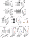
Fig. 6. MT1-MMP cleaves GPR158 to suppress OCN signaling.
a, b Western blotting analysis on the expression of GPR158 in the hippocampus lysates from WT, Mmp14−/− and Mmp14+/− mice (a). Quantification of GPR158 expression relative to β-actin loading control (b). One-way ANOVA followed by Tukey’s multiple comparisons test (n = 4 mice per group). c, d The protein expression of GPR158 in primary hippocampal neurons obtained from WT and Mmp14−/− mice and further treated with either WT MT1-MMP or catalytic inactive MT1-EA (c). Quantification of GPR158 expression relative to β-actin loading control (d). One-way ANOVA followed by Tukey’s multiple comparisons test (n = 3 mice per group). e, f Western blot analyses of GPR158 and MT1-MMP in hippocampus lysates from young and aged WT and Mmp14+/− mice (e) and quantification (f). One-way ANOVA followed by Tukey’s multiple comparisons test (n = 4 mice per group). g, h Representative immunoblots comparing hippocampal GPR158 protein levels in young and aged mice injected with shRNA targeting Mmp14 (shMMP14) or scrambled shRNA (shControl) (g) and quantification of GPR158 expression (h). One-way ANOVA followed by Tukey’s multiple comparisons test (n = 4 mice per group). il In vitro cleavage assay in HEK293 cells expressing human GPR158. Western blot analyses of GPR158 and MT1-MMP in cell lysates transfected with either WT MT1-MMP or catalytic inactive MT1-EA (i). Quantification of the immunoblot for GPR158 expression (j). One-way ANOVA followed by Tukey’s multiple comparisons test (n = 3 mice per group). rGPR158 was incubated with rMT1 from three different sources (in-house, Abcam, and Enzo) and examined for GPR158 and MT1-MMP expression using western blotting assay (k). IP3 accumulation in WT and Mmp14+/− mice hippocampal neurons after 1 h treatment with OCN or glutamate (l). One-way ANOVA followed by Tukey’s multiple comparisons test (n = 5 mice per group). m Schematic depicting bilateral injection of AAV expressing either AAV-WT MT1-MMP, or AAV-MT1-EA followed by OCN injection after two weeks. n The object location task to examine the preference for the displaced object. One-way ANOVA followed by Tukey’s multiple comparisons test (n = 7 for AAV shControl + vehicle, AAV shControl + OCN, AAV-WT MT1-MMP + vehicle, AAV-WT MT1-MMP + OCN, AAV-MT1-EA + OCN and n = 5 mice for AAV MT1-EA + vehicle). oq Percentage of total time spent in the target quadrant (o) and distance traveled to the target hole (p) in the Barnes maze test. One-way ANOVA followed by Tukey’s multiple comparisons test (n = 6 mice per group). Percentage of freezing time during a contextual fear conditioning test (q). One-way ANOVA followed by Tukey’s multiple comparisons test (n = 7 mice per group). Data are expressed as mean ± SEM, and each data point represents individual mouse.
Fig. 7
Fig. 7. MT1-MMP regulates cognitive aging by suppressing Ocn/Gpr158 signaling axis.
a The preference for the displaced object in the object location task by 2-month-old (young), 18-month-old (aged) WT and Mmp14+/− aged mice injected with either shControl or shGpr158. One-way ANOVA followed by Tukey’s multiple comparisons test (n = 7 young + shControl, n = 6 aged + shControl, n = 6 aged + shGpr158, n = 7 Mmp14+/− aged + shControl, n = 6 Mmp14+/− aged + shGpr158). b, c Percentage of total time spent in the target quadrant (b) and distance traveled to the target hole (c) in Barnes maze test. One-way ANOVA followed by Tukey’s multiple comparisons test (n = 6 mice per group). d, e LTP analysis in the CA1 hippocampus region by measuring the fEPSP (d) and the average of fEPSP slope (e). One-way ANOVA followed by Fisher’s LSD post hoc test (n = 5 mice per group). Data are expressed as mean ± SEM, and each data point represents individual mouse.

References

    1. Wyss-Coray, T. Ageing, neurodegeneration and brain rejuvenation. Nature539, 180–186 (2016). - PMC - PubMed
    1. Harry, G. J. Microglia during development and aging. Pharm. Ther.139, 313–326 (2013). - PMC - PubMed
    1. Mosher, K. I. & Wyss-Coray, T. Microglial dysfunction in brain aging and Alzheimer’s disease. Biochem Pharm.88, 594–604 (2014). - PMC - PubMed
    1. Safaiyan, S. et al. Age-related myelin degradation burdens the clearance function of microglia during aging. Nat. Neurosci.19, 995–998 (2016). - PMC - PubMed
    1. Hedden, T. & Gabrieli, J. D. Insights into the ageing mind: a view from cognitive neuroscience. Nat. Rev. Neurosci.5, 87–96 (2004). - PubMed

LinkOut - more resources