. 2025 Oct 7;19(39):34575-34595.
doi: 10.1021/acsnano.5c06003. Epub 2025 Sep 23.

Hydrogel Loaded with Aminoethyl Anisamide-Modified Exosomes Attenuates Hepatic Fibrosis by Targeting Activated Hepatic Stellate Cells

Affiliations

Hydrogel Loaded with Aminoethyl Anisamide-Modified Exosomes Attenuates Hepatic Fibrosis by Targeting Activated Hepatic Stellate Cells

Zongbin Sun et al. ACS Nano. .

Abstract

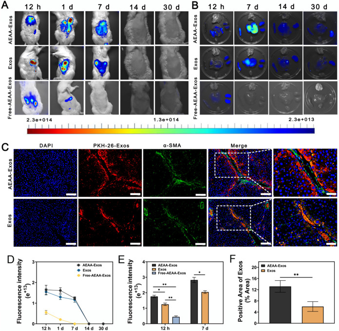
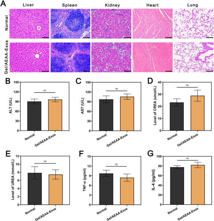
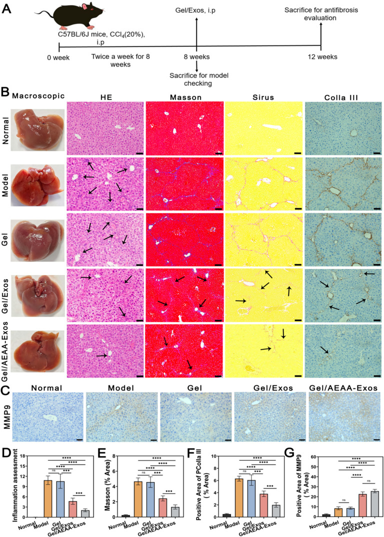
Stem cell-based regenerative research has highlighted the therapeutic potential of human umbilical cord mesenchymal stem cell-derived exosomes (hucMSC-Exos) for hepatic tissue regeneration and repair. However, exosomes undergo rapid clearance following systemic administration, limiting their therapeutic potential because of insufficient retention and sustained release. In this study, an innovative hydrogel-mediated delivery platform encapsulating aminoethyl anisamide (AEAA)-functionalized exosomes was developed to mitigate hepatic fibrosis. By synthesizing a hydrogel (CMC-OD/TA-Fe(III), Gel) composed of carboxymethyl chitosan, oxidized dextran, and iron tannate, and then encapsulating umbilical cord mesenchymal stem cell-derived exosomes functionalized by AEAA (AEAA-Exos), we implanted this Gel/AEAA-Exos into mice with hepatic fibrosis by intraperitoneal injection to evaluate the therapeutic effect of the hydrogel. The hydrogel had favorable physical properties, optimal biocompatibility, and a sustained-release profile. And Gel/AEAA-Exos system significantly reduced oxidative stress and alleviated hepatic fibrosis. Additionally, RNA-seq revealed that the Gel/AEAA-Exos system ameliorates hepatic fibrogenesis mainly by modulating oxidative stress, collagen deposition, and inflammatory cascade in liver tissues. This strategy offers a targeted and efficient approach for treating liver fibrosis induced by chronic hepatic injury and improves targeting efficiency and therapeutic outcomes through engineered exosome delivery.

Keywords: AEAA-engineered exosomes; antihepatic fibrosis; hydrogel; sustained release; target.


Figures

1
1
Evaluation of exosomes and hydrogel properties. (A) Transmission electron microscopy (TEM) imaging was employed to characterize the ultrastructural features of exosomes (scale bar: low: 200 nm, high: 100 nm). (B) Nanoscale exosomes sizing was performed via Nanoparticle Tracking Analysis (NTA) capturing Brownian motion trajectories. (C) Exosomal biomarkers (TSG101, CD9, CD89) were quantified via Western blotting to validate vesicle identity. (D) Micromorphology analysis of different hydrogels after freeze-drying by SEM (scale bar: low: 100 μm, high: 50 μm). (E) SEM microstructure of CMC-OD/TA-Fe/AEAA-Exos hydrogels and morphology of exosomes attached to the surface of hydrogels at high magnification (scale bar: low: 50 μm, high:1 μm). (F) Elemental energy spectrum analysis of CMC-OD/TA-Fe/AEAA-Exos hydrogels (scale bar: 250 μm). (n = 3).
1
1. Based on Schiff Base Reaction, a New Hydrogel CMC-OD/TA-Fe­(III) Was Prepared by CMC, OD and TA-Fe­(III)
2
2
Characterization of hydrogels’ synthesis and properties. (A) Fourier-transform infrared spectroscopy of hydrogel components. (B) 1H NMR assay of dextran and oxidized dextran. (C, D) show the colloidal states of CMC-OD and CMC-OD/TA-Fe hydrogels, respectively. (E) Injectable ability test of CMC-OD/TA-Fe hydrogel. (F) Test of self-healing properties for CMC-OD/TA-Fe hydrogel. (G) Viscoelastic test of CMC-OD/TA-Fe hydrogel. (H) The adhesion properties of CMC-OD and CMC-OD/TA-Fe hydrogels were evaluated by porcine skin tests. (I) Analysis of degradation characteristics of hydrogels. (J) Analysis of swelling properties of hydrogels. (K) Rheological properties of CMC-OD/TA-Fe hydrogel were evaluated by angular frequency test, stress test and stepwise strain test, respectively. Data are presented as means ± SD (n = 3).
3
3
In vitro biosafety assessment of hydrogel/exosome systems. (A) Live and dead stain results of L929 cells treated with different groups of hydrogel extracts (scale bar: 100 μm). (B) Adhesion tests of L929 cells and BRL-3A cells grown on different hydrogel surfaces (scale bar: 100 μm). (C) Quantification analysis of the live L929 cell-positive expression area by ImageJ software correspond to (A). (D) Quantification analysis of the live L929 and BRL-3A cells-positive expression area by ImageJ software correspond to (B). (E) CCK-8 cytotoxicity assay of hydrogels on L929 cells. (F) CCK-8 cytotoxicity assay of hydrogels on BRL-3A cells. (G) CCK-8 cytotoxicity assay of different hydrogels on BRL-3A cells by CCK-8 cytotoxicity assay. (H) Assessment for the effect of hydrogels on hemolysis. (I, J) Assessment for the effects of hydrogel coagulation. Data are presented as means ± SD (n = 3). P > 0.05 (ns, not significant).
4
4
Evaluation of biological function of hydrogel/exosome systems in vitro. (A) Evaluation of targeting of activated hepatic stellate cells by gel/AEAA-Exos systems (scale bar: 50 μm). (B) Evaluation of anti-ROS ability in gel/AEAA-Exos systems (scale bar: 400 μm). (C) The antifibrotic effect of gel/AEAA-Exos systems was evaluated by α-SMA index (scale bar: 100 μm). (D) The antifibrosis effect of gel/AEAA-Exos systems was evaluated by Colla I index (scale bar: 100 μm). Quantification analysis of exosomes-positive area­(E), DCFH-positive area (F), α-SMA-positive area (G) and Colla I-positive area (H). Data are presented as means ± SD (n = 3). **P < 0.01, ***P < 0.001, ****P < 0.0001.
5
5
Evaluation of in vivo targeting ability of hydrogel/exosome systems. (A) Small animals imaging results of after free AEAA-exosomes (Free-AEAA-Exos group), gel/Exos (Exos group) and gel/AEAA-Exos (AEAA-Exos group) systems implantation in SD rats at different observation points (12 h, 1 day, 7 days, 14 days, 30 days). (B) Evaluation of the distribution of exosomes in the liver after free AEAA-exosomes, gel/Exos and gel/AEAA-Exos systems implantation in SD rats at different times (12 h, 7 days, 14 days, 30 days). (C) After gel/PKH-26-Exos system implantation in SD rats for 7 days, the targeting performance of exosomes on activated hepatic stellate cells was evaluated by tissue immunofluorescence assay (scale bar: low: 100 μm, high: 40 μm). (D) Fluorescence quantitative analysis of the distribution of exosomes in SD rats in vitro corresponding to (A). (E) Fluorescence quantitative analysis of the distribution of exosomes in the liver of SD rats in vivo corresponding to (B). (F) Quantification analysis of the distribution of exosomes in the liver of SD rats corresponding to (C). Data are presented as means ± SD (n ≥ 3). *P < 0.05,**P < 0.01.
6
6
Biosafety evaluation of hydrogel/exosome systems in vivo. (A) HE stain pictures of liver, spleen, kidney, heart and lung of Normal group and experimental group after gel/AEAA-Exos implantation in mice for 7 days (scale bar: 400 μm). (B–E) evaluated the biosafety of gel/AEAA-Exos systems was evaluated by serum ALT, AST, CREA and UREA levels, respectively. (F, G) The biosafety of gel/AEAA-Exos system was evaluated by ELISA kit for serum TNF-α and IL-4 levels, respectively. (n = 5). P > 0.05 (ns, not significant).
7
7
Evaluation of in vivo antifibrosis ability of hydrogel/exosome systems. (A) The process of establishing hepatic fibrosis modeling and antihepatic fibrosis is shown. (B) To evaluate the antifibrosis effect of gel/AEAA-Exos systems by HE, Masson, sirius red stain and PColla III (scale bar: 200 μm). (C) The antifibrosis effect of gel/AEAA-Exos system was evaluated by MMP9 index (scale bar: 200 μm). Quantification analysis of inflammation-positive area (D), Masson-positive area (E), PColla III-positive area (F) and MMP9-positive area (G). Data are presented as means ± SD (n = 5). ***P < 0.001, ****P < 0.0001, P > 0.05 (ns, not significant).
8
8
Immunofluorescence assessment and serological assessment of in vivo antihepatic fibrosis of the hydrogel/exosome system. (A, B) The antifibrosis effect of gel/AEAA-Exos system was evaluated by TGF-β and α-SMA, respectively (scale bar of A: low: 100 μm, high: 20 μm, scale bar of B: low: 50 μm, high: 20 μm). (C) Quantification analysis of the TGF-β-positive expression area, the α-SMA-positive expression area. (D) Oxidative stress levels (SOD and MDA) evaluated by Elisa. (E) Serum ALT and AST levels evaluated by Elisa. (F) Inflammatory levels (TNF-α and IL-4) evaluated by Elisa. Data are presented as means ± SD (n = 5). **P < 0.01, ***P < 0.001, ****P < 0.0001, P > 0.05 (ns, not significant).
9
9
Transcriptome RNA sequencing to evaluate the mechanism of Gel/AEAA-Exos delivery system in alleviating liver fibrosis. (A) Venn diagram of different groups of differential genes. (B, C) Volcanic maps of differential genes in model group and Gel/AEAA-Exos group. (D, E) KEGG Scatter Plot analysis of differential gene-related pathways in the model group and Gel/AEAA-Exos group. (F) Heat map analysis of genes related to antifibrosis pathways in this study. (G) The gene expression levels of MAP3K13, ASK1, MAPK14, MAPK1, HSPB1 and MMP9 were evaluated by RT-PCR. Data are presented as means ± SD (n = 3). *P < 0.05, **P < 0.01, ***P < 0.001.

References

    1. Dhar D., Baglieri J., Kisseleva T., Brenner D. A.. Mechanisms of liver fibrosis and its role in liver cancer. Exp. Biol. Med. 2020;245:96–108. doi: 10.1177/1535370219898141. - DOI - PMC - PubMed
    1. Parola M., Pinzani M.. Liver fibrosis: Pathophysiology, pathogenetic targets and clinical issues. Mol. Aspects Med. 2019;65:37–55. doi: 10.1016/j.mam.2018.09.002. - DOI - PubMed
    1. Caligiuri A., Gentilini A., Pastore M., Gitto S., Marra F.. Cellular and Molecular Mechanisms Underlying Liver Fibrosis Regression. Cells. 2021;10:2759. doi: 10.3390/cells10102759. - DOI - PMC - PubMed
    1. Llovet J. M., Kelley R. K., Villanueva A., Singal A. G., Pikarsky E., Roayaie S., Lencioni R., Koike K., Zucman-Rossi J., Finn R. S.. Hepatocellular carcinoma. Nat. Rev. Dis Primers. 2021;7:6. doi: 10.1038/s41572-020-00240-3. - DOI - PubMed
    1. Gines P., Krag A., Abraldes J. G., Sola E., Fabrellas N., Kamath P. S.. Liver cirrhosis. Lancet. 2021;398:1359–1376. doi: 10.1016/S0140-6736(21)01374-X. - DOI - PubMed