. 2025 Sep 30;122(39):e2504697122.
doi: 10.1073/pnas.2504697122. Epub 2025 Sep 23.

ALKBH5 demethylates the m6A modification of SOCS3 in microglia/macrophages and alleviates neuroinflammation after brain injury

Affiliations

ALKBH5 demethylates the m6A modification of SOCS3 in microglia/macrophages and alleviates neuroinflammation after brain injury

Lin Cai et al. Proc Natl Acad Sci U S A. .

Abstract

Microglia/macrophage-induced neuroinflammation plays a crucial role in the progression of traumatic brain injury (TBI). However, the involvement of N6-methyladenosine (m6A) RNA modifications in this process remains elusive. Single-cell RNA sequencing (scRNA-seq) and m6A RNA immunoprecipitation sequencing (MeRIP-seq) across multiple time points postinjury revealed a strong correlation between m6A modifications and genes enriched in microglia/macrophages. Furthermore, the m6A demethylase ALKBH5 was identified as a key regulator of dynamic m6A patterns at the injury site. ALKBH5 suppression in microglia/macrophages exacerbated neuroinflammation in vitro and worsened neurological deficits in controlled cortical impact (CCI) models. MeRIP-qPCR and RNA pull-down assays revealed SOCS3 was a downstream target of ALKBH5-mediated m6A demethylation. This demethylation stabilized Socs3 mRNA and enhanced its protein expression, which in turn suppressed neuroinflammation via inhibiting the JAK2-STAT3 pathway. Conversely, SOCS3 depletion impaired functional recovery after injury. These findings unveiled a critical ALKBH5-m6A-SOCS3 regulatory axis that mitigated microglia/macrophage-driven neuroinflammation after TBI, underscoring its potential as a therapeutic intervention target for TBI progression.

Keywords: ALKBH5; N6-methyladenosine; SOCS3; microglia; neuroinflammation.


Conflict of interest statement

Competing interests statement:The authors declare no competing interest.

Figures

Fig. 1.
Fig. 1.
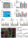
Microglia/macrophages in injured cortical tissues played a key role in regulating pathological process of TBI. (A) tSNE plots of scRNA-seq in injured cortical tissue at different time points postinjury. (B) Expression of TBI-related pathological biomarkers during acute and subacute phases postinjury in microglia/macrophages.
Fig. 2.
Fig. 2.
Conjoint analysis of m6A-seq with transcriptome RNA-seq in mice CCI models of injured cortical tissues. (A) Schematic representation of sample preparation process and mechanisms for m6A-seq analysis. (B) Metagene profiles showing the distribution of m6A peaks at different time points postinjury in IP and input groups. (C) IP/input m6A peak density across the 5′-UTR, CDS, and 3′-UTR, arrows indicated locations where m6A peaks changed. (D) Sequence motif was identified within m6A peaks by HOMER analysis. (E) Four-quadrant analysis of differentially expressed genes (DEGs) and their m6A modification levels compared with sham group. Gene log2 (FC) represented the fold change of gene expression at the transcriptomic level, while m6A Diff. log2 (FC) reflected the changes in m6A modification for the same genes. Statistical criteria were fold changes in gene expression and m6A modification levels ≥2, with P < 0.05 compared with the sham group. Yellow indicated genes with increased expression and decreased m6A modification levels; blue indicated increased expression and increased m6A levels; green indicated decreased expression and decreased m6A levels; purple indicated decreased expression and increased m6A levels.
Fig. 3.
Fig. 3.
The demethylase ALKBH5 might regulate the pathological progression of injured cortical tissues in mice CCI models by modulating m6A levels in microglia/macrophages. (A) Dot blot analysis showing m6A modification levels in transcriptome of injured cortical tissue at different time points postinjury, statistical results are shown in Right panel. N = 3/group. (B) Western blot results of ALKBH5 expression in injured cortical tissue, GAPDH was used as loading control. N = 2 in sham group and N = 3 in TBI groups. (C) Correlation analysis between ALKBH5 protein expression and m6A modification levels. (D) Ridge plots from scRNA-seq data revealed that Alkbh5 expression in microglia/macrophages showed a significant temporal correlation. Alkbh5 expression in different cell types was shown in tSNE plots. Data were presented as means ± SEMs. Student’s t test, ***P < 0.001; **P < 0.01; *P < 0.05 vs Sham group.
Fig. 4.
Fig. 4.
The demethylase ALKBH5 might inhibit microglia-mediated neuroinflammation by regulating m6A levels. (A and B) After cotreatment with siAlkbh5 and LPS for 36 h, BV2 cells (100 ng/ml LPS), mouse, and rat primary microglia cells (50 ng/ml LPS) were subjected to immunoblot analysis for ALKBH5 expression and ACTIN was used as loading control (A), statistical results are shown in Fig. 4B. (C and D) Dot blot detection of m6A modification levels in BV2 cell lines, rat, and mouse primary microglia (C), statistical results are shown in Fig. 4D. (EG) qPCR analysis of proinflammatory cytokine mRNA expression levels in three microglia cell types. N = 3/group. (H and I) Representative fluorescence images showing dual staining of ALKBH5 (green) with microglia-specific biomarker IBA1 (red) (H), statistical results of ALKBH5 distribution patterns across experimental conditions are shown in Fig. 4I. Cell nuclei are shown in blue with DAPI. Scale bar, 20 μm for BV2 cells and 25 μm for primary microglia cells. Cells without LPS and siAlkbh5 treatment were shown as Control. Data were presented as means ± SEMs. One-way ANOVA with Tukey’s post hoc test, ***P < 0.001; **P < 0.01; *P < 0.05 vs Control group. ###P < 0.001; ##P < 0.01; #P < 0.05 vs LPS group.
Fig. 5.
Fig. 5.
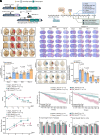
Microglia-specific knockout of ALKBH5 exacerbated neurological damage and delayed functional motor recovery in mice CCI models. (A) Breeding strategies for microglia-specific Alkbh5 conditional knockout (cKO) mice. For negative control, CreER allele-deficient (Alkbh5Ctrl) offspring mice were selected. (B) Overview of experimental timeline for in vivo studies and neurobehavioral assessments. (C) Comparison of cortical tissue damage in mice at different time points after CCI modeling. (D and E) Gross pathological changes in consecutive coronal brain sections stained with cresyl violet at indicated time points postinjury, the representative images were selected to illustrate the general trend (D), the percentage of brain tissue loss are shown in Fig. 5E. N = 8/group. (F and G) Representative images of EB extravasation in groups at indicated time points postinjury. Blue area represented the site of dye extravasation (F). Statistical results are shown in Fig. 5G. N = 8/group. (HK) Behavioral function assessments at different time points after CCI modeling, including mNSS scores (H), hanging wire test (I), rotarod test (J), and grip strength test (K). Data were presented as mean ± SEMs. Student’s t test.
Fig. 6.
Fig. 6.
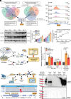
ALKBH5 might be involved in regulating m6A modification of SOCS3. (A) Venn diagram analysis of 62 candidate genes from m6A-seq data with transcriptome datasets from NCBI public databases (GSE180811, GSE58484, GSE44625). (B) IGV browser tracks showing that m6A peaks were enriched in 3’-UTR of Socs3 transcript. (CE) Western blot detection of ALKBH5 and SOCS3 protein levels in sham and injured cortical tissue, GAPDH was used as loading control (C). Statistical results showed the changes in ALKBH5 expression corresponded with the differences in SOCS3 protein expression (D), correlation analysis are shown in Fig. 6E. N = 2 in the sham group and N = 3 in TBI groups. (F and G) Regulating ALKBH5 expression in BV2 and rat primary microglia cells, then subjected cells to MeRIP-qPCR analysis to further detect the level changes in Socs3-m6A at different predicted modification sites. N = 3/group. (H and I) Proteins pulled down by the probe in BV2 cells were detected through western blot. Workflow illustrated in Fig. 6H and probes were constructed for murine-derived Socs3-m6A modification sites. Proteins precipitated by probe in BV2 cells were detected using an anti-ALKBH5 primary antibody (I). Input referred to whole protein lysates from cells and GAPDH as loading control. Data were presented as means ± SEMs. Student’s t test, ***P < 0.001; **P < 0.01; *P < 0.05 vs Sham group.
Fig. 7.
Fig. 7.
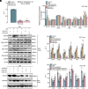
ALKBH5 restrained microglia-mediated neuroinflammation via regulating SOCS3 expression. (A) Cells were treated with siSocs3 for 36 h in BV2 cells, then the knockdown efficiency was validated by qPCR. N = 3/group. (B and C) After cotreatment with siSocs3 and LPS for 36 h, BV2 cells (100 ng/ml LPS) were subjected to immunoblot analysis for ALKBH5, Total/p-STAT3, Total/p-JAK2, Total/p-NFκB expression, ACTIN was used as loading control (B), statistical results are shown in Fig. 7C. (D) qPCR analysis of proinflammatory cytokines mRNA expression levels in BV2 cells. N = 3/group. (E) Lentiviral transfection was used to up-regulate ALKBH5 protein expression and then down-regulating SOCS3 expression via using siRNA. Transfection efficiency was verified by western blot and ACTIN as loading control. (F) qPCR analysis of proinflammatory cytokines mRNA expression levels in BV2 cells. Cells without treatment were shown as Control. N = 3/group. Data were presented as means ± SEMs. One-way ANOVA with Tukey’s post hoc test, ***P < 0.001; **P < 0.01; *P < 0.05 vs Control group. ###P < 0.001; ##P < 0.01; #P < 0.05 vs LPS group.

References

    1. Smith D. H., Meaney D. F., Shull W. H., Diffuse axonal injury in head trauma. J. Head Trauma Rehabil. 18, 307–316 (2003). - PubMed
    1. Humble S. S., et al. , Prognosis of diffuse axonal injury with traumatic brain injury. J. Trauma Acute Care Surg. 85, 155–159 (2018). - PMC - PubMed
    1. Xu X., et al. , Anti-inflammatory and immunomodulatory mechanisms of atorvastatin in a murine model of traumatic brain injury. J. Neuroinflammation 14, 167 (2017). - PMC - PubMed
    1. Hanisch U.-K., Kettenmann H., Microglia: Active sensor and versatile effector cells in the normal and pathologic brain. Nature neuroscience 10, 1387–1394 (2007). - PubMed
    1. Panday A., et al. , Transcription factor NF-κB: An update on intervention strategies. Arch. Immunol. Ther. Exp. 64, 463–483 (2016). - PubMed

MeSH terms

LinkOut - more resources