

Novel insights into the relationship between glaucoma and brain diseases from the genetic to diseases levels: A cross-sectional study
- PMID: 40988281
- PMCID: PMC12459570
- DOI: 10.1097/MD.0000000000044416
Novel insights into the relationship between glaucoma and brain diseases from the genetic to diseases levels: A cross-sectional study
Abstract
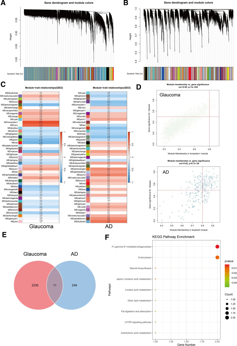
Glaucoma is a heterogeneous group of diseases which is one of the leading causes of irreversible blindness worldwide. Although the eye-brain axis has been proposed, its functional connectivity remains poorly defined. This study aimed to explore the mechanisms and causal relationship between glaucoma and brain cortical structure, focusing on the eye-brain axis. A Mendelian randomization (MR) study was conducted using inverse variance weighting as the primary estimator, alongside MR-PRESSO, MR-Egger, and weighted median methods to assess sensitivity, heterogeneity, and pleiotropy. Pathway analysis, transcriptomic analysis, and weighted gene co-expression network analysis (WGCNA) were applied to investigate brain-eye interactions in Alzheimer's disease (AD) and primary open-angle glaucoma (POAG), revealing shared pathogenic mechanisms. Significant associations between glaucoma and brain cortex regions, including the superior temporal sulcus, anterior cingulate, cuneus, entorhinal, inferior temporal, and insula, were identified. About 18 overlapping genes between AD and POAG were found, including MYH14, EFNA1, FZD1, and CACNG3. Using WGCNA, 11 overlapping genes were identified as most related to both AD and POAG, including TSC2, MAGED4, LSS, and DNM1. These results contributed to understanding the association between glaucoma and the brain, indicating the eye-brain axis and may provide clues for early screening of high-risk populations.
Keywords: Alzheimer’s disease; IOP; Mendelian randomization; brain cortical structure; glaucoma; visual field defects.
Copyright © 2025 the Author(s). Published by Wolters Kluwer Health, Inc.
Conflict of interest statement
The authors have no conflicts of interest to disclose.
Figures


References
-
- GBD 2019 Blindness and Vision Impairment Collaborators; Vision Loss Expert Group of the Global Burden of Disease Study. Causes of blindness and vision impairment in 2020 and trends over 30 years, and prevalence of avoidable blindness in relation to VISION 2020: the right to sight: an analysis for the global burden of disease study. Lancet Glob Health. 2021;9:e144–60.
-
- Casson RJ, Chidlow G, Wood JP, Crowston JG, Goldberg I. Definition of glaucoma: clinical and experimental concepts. Clin Exp Ophthalmol. 2012;40:341–9. - PubMed
-
- Tham YC, Li X, Wong TY, Quigley HA, Aung T, Cheng C-Y. Global prevalence of glaucoma and projections of glaucoma burden through 2040: a systematic review and meta-analysis. Ophthalmology. 2014;121:2081–90. - PubMed
MeSH terms
Grants and funding
LinkOut - more resources
Full Text Sources
Medical
Miscellaneous
