

Metastatic Retroperitoneal Leiomyosarcoma to the Right Lower Eyelid Presenting as a Chalazion
- PMID: 40988997
- PMCID: PMC12438942
- DOI: 10.18502/jovr.v20.16421
Metastatic Retroperitoneal Leiomyosarcoma to the Right Lower Eyelid Presenting as a Chalazion
Abstract
Purpose: Leiomyosarcoma (LMS) is an aggressive tumor with a high metastatic rate that rarely metastasizes to the periocular region.
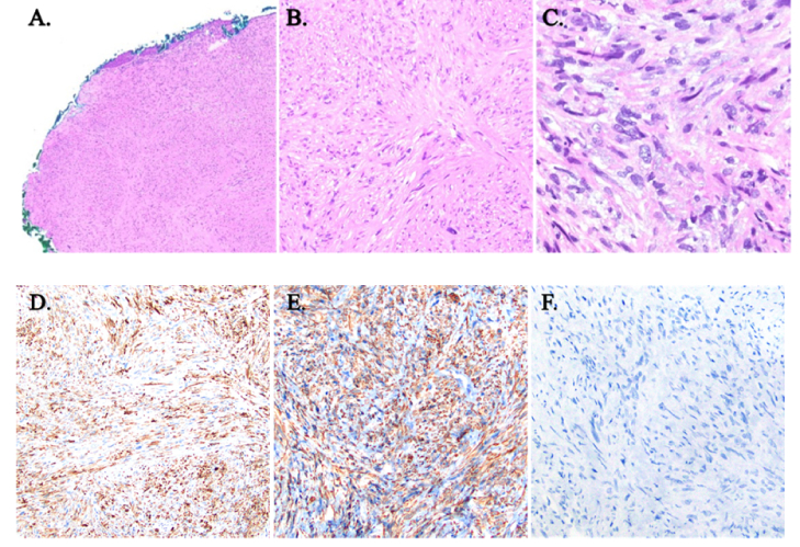
Methods: A 50-year-old male with a previous two-year history of primary stage IV LMS presented with metastatic retroperitoneal LMS, which was initially incorrectly described as an eyelid chalazion refractory to medical management. An excisional biopsy sent to pathology revealed metastatic retroperitoneum LMS. There was resolution of ocular irritation following biopsy, and an oncology referral was made.
Conclusion: This case of metastatic LMS to the eyelid mimicking a chalazion is rare, as only six other cases have been described previously. Our case contributes to this discussion by highlighting the importance of considering metastatic disease and performing a full-thickness biopsy in a patient presenting with a non-resolving eyelid chalazion. Recognizing tumor spread to the eyelid can be an important step in the diagnosis, surveillance, and management of metastatic LMS.
Keywords: Leiomyosarcoma; Chalazion; Eyelid; Metastatic; Oculoplastic Surgery.
Copyright © 2025 Eid et al.
Conflict of interest statement
None.
Figures

References
-
- Campanacci M. Campanacci M, editor. Bone and soft tissue tumors: Clinical features, imaging, pathology and treatment. Springer; Leiomyosarcoma; 1999 pp.
-
- Toro JR, Travis LB, Wu HJ, Zhu K, Fletcher CD, Devesa SS. Incidence patterns of soft tissue sarcomas, regardless of primary site, in the surveillance, epidemiology and end results program, 1978-2001: An analysis of 26,758 cases. Int J Cancer. 2006;119:2922–2930. - PubMed
-
- White VA, Damji KF, Richards JS, Rootman J. Leiomyosarcoma of the conjunctiva. Ophthalmology. 1991;98:1560–1564. - PubMed
-
- Voros GM, Birchall D, Ressiniotis T, Neoh C, Owen RI, Strong NP. Imaging of metastatic orbital leiomyosarcoma. Ophthalmic Plast Reconstr Surg. 2005;21:453–455. - PubMed
Publication types
LinkOut - more resources
Full Text Sources