Case Reports
. 2025 Sep 8:15:1608291.
doi: 10.3389/fonc.2025.1608291. eCollection 2025.

Case report series: Pembrolizumab and enfortumab vedotin in plasmacytoid urothelial carcinoma

Affiliations
Case Reports

Case report series: Pembrolizumab and enfortumab vedotin in plasmacytoid urothelial carcinoma

Susanna G Bowen et al. Front Oncol. .

Abstract

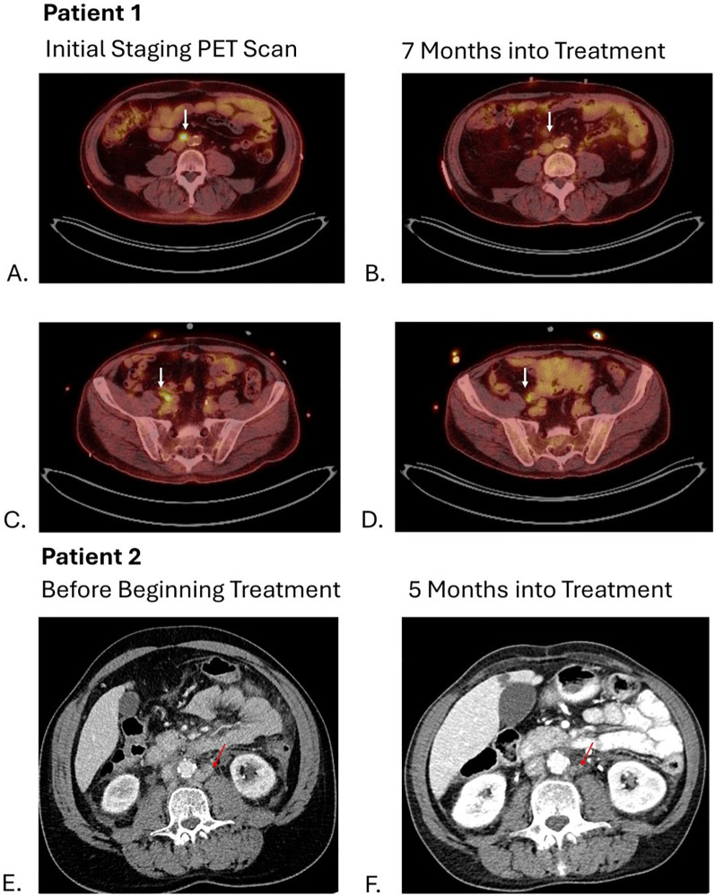
Plasmacytoid urothelial carcinoma (PUC) is a rare and aggressive histologic subtype of urothelial carcinoma with no well-established treatment. Recently, the combination of pembrolizumab and enfortumab vedotin has become the standard of care for locally advanced and metastatic urothelial carcinoma due to improved survival outcomes in the EV-302 trial, but the number of histological subtypes in this trial is unknown. This case series presents three patients with Stage IV PUC who were treated with the combination of pembrolizumab and enfortumab vedotin. Two of the three patients demonstrated sustained stable disease after eight and ten months of treatment with this combination with manageable adverse effects including rash and colitis. The third patient experienced disease progression to leptomeningeal involvement eight months following initial diagnosis and subsequently succumbed to the disease. These observations support the potential efficacy of pembrolizumab in combination with enfortumab vedotin as a therapeutic option for this aggressive urothelial carcinoma subtype.

Keywords: antibody drug conjugate; enfortumab vedotin; immune-checkpoint inhibitors; pembrolizumab; plasmacytoid; urothelial carcinoma.


Conflict of interest statement

The authors declare that the research was conducted in the absence of any commercial or financial relationships that could be construed as a potential conflict of interest.

Figures

Figure 1
Figure 1
Slides (A, B) represent patient 1. (A) Muscle-invasive plasmacytoid urothelial carcinoma. Discohesive single individual tumor cells are seen diffusely infiltrating around and through muscular fascicles. The tumor cells display eccentric nuclei with peripheral eosinophilic cytoplasm, H&E stain, 20x magnification. (B) Large muscular fascicles are seen with tumor cells infiltrating through fascial planes, which is commonly seen in plasmacytoid urothelial carcinoma. The tumor cells show eccentric nuclei with peripheral eosinophilic cytoplasm, as well as signet ring cell forms, which are characteristic of plasmacytoid urothelial carcinoma, H&E stain, 20x magnification. Slides C and D represent patient 2. (C) Muscle-invasive plasmacytoid urothelial carcinoma. Tumor cells are seen in single file lines and are diffusely infiltrative through a myxoid stroma. Note the presence of an atypical mitotic figure, as well as scattered plasmacytoid and signet ring cell forms, H&E stain, 20x magnification. (D) Note the infiltration through and around muscular fascicles, as well as the abundant mitotic figures, H&E stain, 20x magnification. Slides E-H represent patient 3. (E) Invasive plasmacytoid urothelial carcinoma involving the lamina propria. The sample is superficial sample with no muscularis propria identified; however, the tumor is diffusely infiltrating through lamina propria with many discohesive cells with eccentric nuclei and peripheral eosinophilic cytoplasm, H&E stain, 20x magnification. (F) Nuclear staining is seen with the GATA-3 immunostain, GATA-3 stain, 20x magnification. (G) Membranous and cytoplasmic staining is seen with the CK7 immunostain, CK7 stain, 20x magnification. (H) E-cadherin staining is lost in the tumor cells, which is common in plasmacytoid urothelial carcinoma, E-cadherin stain, 20x magnification.
Figure 2
Figure 2
Arrows on images (A, C) show hypermetabolic activity of aortocaval and right common iliac lymph nodes respectively with decrease in metabolic activity shown 7 months into treatment on images (B, D). Arrows on images (E, F) show decrease in size of left para-aortic lymph nodes from 11 mm to 3 mm.

References

    1. Li Q, Assel M, Benfante NE, Pietzak EJ, Herr HW, Donat M, et al. The impact of plasmacytoid variant histology on the survival of patients with urothelial carcinoma of bladder after radical cystectomy. Eur Urol Focus. (2019) 5:104–8. doi: 10.1016/j.euf.2017.06.013, PMID: - DOI - PMC - PubMed
    1. Benabdallah W, Ben Othmane M, Ouahchi I, Mestiri S, Belkacem O, Bouassida K, et al. Plasmacytoid bladder cancer: a rare case report. Ann Med Surg (Lond). (2023) 85:1885–7. doi: 10.1097/MS9.0000000000000374, PMID: - DOI - PMC - PubMed
    1. Kim DK, Kim JW, Ro JY, Lee HS, Park JY, Ahn HK, et al. Plasmacytoid variant urothelial carcinoma of the bladder: A systematic review and meta-analysis of clinicopathological features and survival outcomes. J Urol. (2020) 204:215–23. doi: 10.1097/JU.0000000000000794, PMID: - DOI - PubMed
    1. Lobo N, Shariat SF, Guo CC, Fernandez MI, Kassouf W, Choudhury A, et al. What is the significance of variant histology in urothelial carcinoma? Eur Urol Focus. (2020) 6:653–63. doi: 10.1016/j.euf.2019.09.003, PMID: - DOI - PubMed
    1. Sun M, Schaap A, Robinson BD, Nanus DM, Tagawa ST. A case report of a patient with plasmacytoid urothelial cancer with significant response to HER2-targeting therapy and enfortumab vedotin. Am J Clin Exp Urol. (2021) 9:390–6., PMID: - PMC - PubMed

Publication types

LinkOut - more resources