. 2025 Oct:120:105934.
doi: 10.1016/j.ebiom.2025.105934. Epub 2025 Sep 23.

DNA demethylation mediated endogenous retroviruses transcription promotes aberrant T cell differentiation in systemic lupus erythematosus via RIG-I pathway

Affiliations

DNA demethylation mediated endogenous retroviruses transcription promotes aberrant T cell differentiation in systemic lupus erythematosus via RIG-I pathway

Xiaoli Min et al. EBioMedicine. 2025 Oct.

Abstract

Background: Systemic lupus erythematosus (SLE) displays quantitative and/or qualitative deficiencies of regulatory T cells (Treg) and expansion and hyperfunction of pathogenic T cells. However, the underlying mechanism of dysregulated T lymphocyte differentiation in SLE remains unclear.

Methods: Transcriptome sequencing and functional assays were performed to elucidate the mechanisms and function of human endogenous retroviruses (HERVs) on T cell differentiation in SLE. The effect of retinoic acid-inducible gene I (RIG-I) deficiency on lupus pathogenesis were assessed in lupus-like mouse models.

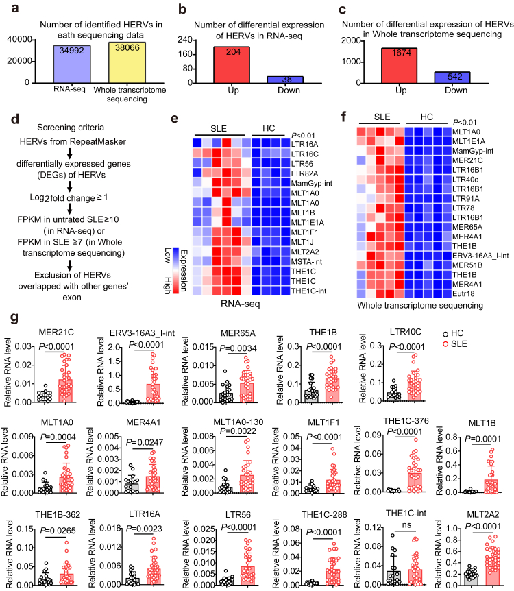
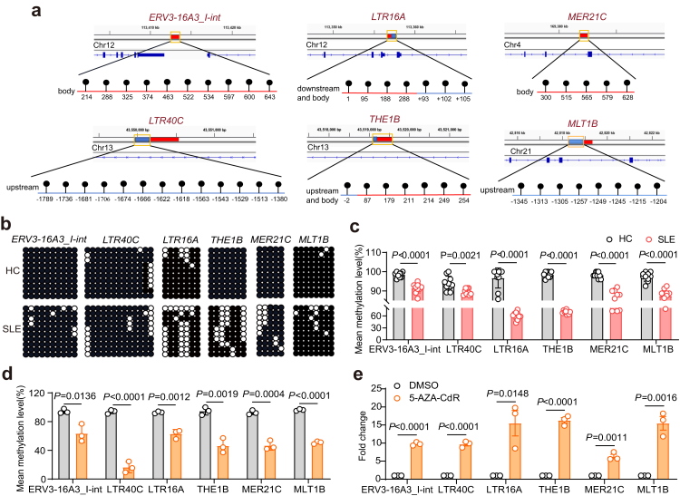
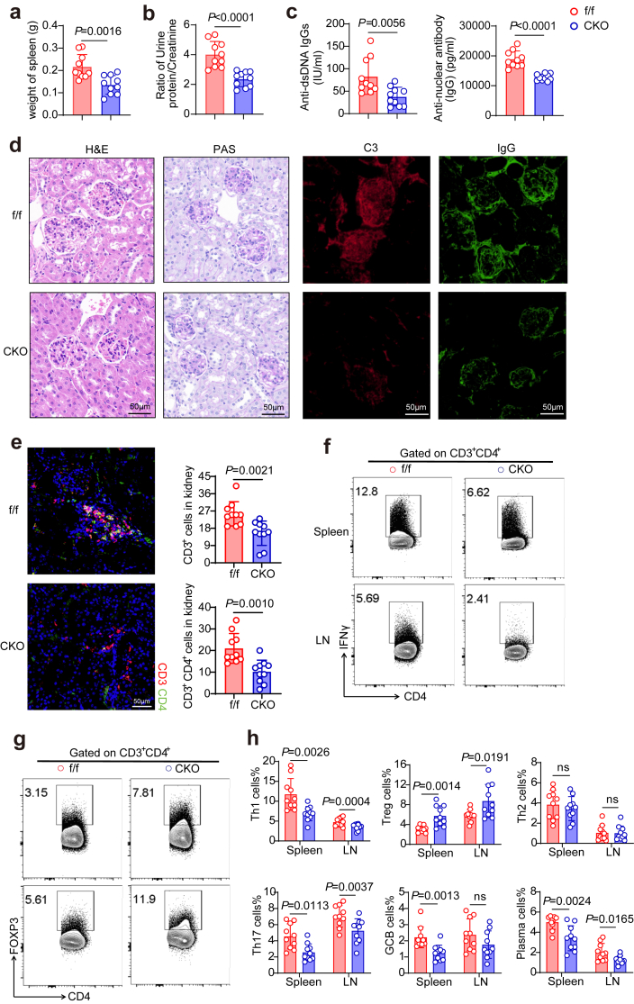
Findings: We found that many transcripts derived from HERVs were highly expressed in CD4+ T cells from patients with SLE due to DNA hypomethylation, some of which were characterized by double strand RNAs (dsRNAs). Excessive dsRNAs promoted Th1 cell differentiation and inhibited Treg cell differentiation via the activation of the dsRNA sensor RIG-I pathway, accompanied by a high level of type I interferons (IFN-I) and interferon-stimulated gene expression. In contrast, T cell-specific ablation of RIG-I alleviated disease progression in lupus-like mouse models by reducing the proportion of pathogenic T cells, restoring the percentage of Treg cells, and diminishing cytokine and autoantibody release. Importantly, we demonstrated that the dsRNA-induced RIG-I/IRF3 pathway regulated Th1 cell differentiation in a IFN-I/STAT1 signalling-dependent manner and inhibited Treg cell differentiation via SMAD3 signalling.

Interpretation: Our findings reveal the roles of HERV-derived dsRNA/RIG-I pathway in regulating the aberrant differentiation of T cells in patients with SLE and may facilitate the development of potential therapeutic targets for SLE.

Funding: A full list of funding sources can be found in the Funding section.

Keywords: Human endogenous retroviruses; RIG-I pathway; Systemic lupus erythematosus; T cell differentiation.


Conflict of interest statement

Declaration of interests The authors declare no competing financial interests.

Figures

Fig. 1
Fig. 1
Screening and identification of the highly expressed HERVs in CD4+ T Cells from patients with SLE. We performed RNA-seq (data included CD4+T cells of 4 healthy controls and 6 patients with SLE) and whole transcriptome sequencing (data included CD4+T cells of 5 healthy controls and 5 patients with SLE). a Numbers of HERVs identified by RNA-seq and whole transcriptome sequencing. b, c Numbers of differential expression of HERVs in RNA-seq and the whole transcriptome sequencing. d Schematic diagram depicting the screening of HERVs in CD4+ T Cells of SLE. FPKM, fragments per kilobase of transcript per million mapped reads. e, f Differential expression of HERVs were identified with significantly higher expression in RNA-seq and the whole transcriptome sequencing according to stringent thresholds. g RT-qPCR analysis of the identified HERVs expression in CD4+ T cells of patients with SLE (n = 30) and HCs (n = 16). (unpaired 2-tailed Student's t test and Mann–Whitney test for eg).
Fig. 2
Fig. 2
Detection of the DNA methylation status of HERV loci in CD4+ T Cells of SLE. a CpG islands in HERV loci (blue bars represent upstream or downstream areas of genes; red bars represent gene body areas; yellow boxes represent the detected CpG regions). b, c DNA methylation levels of HERVs in CD4+ T cells from HCs and patients with SLE (n = 10 each group). d Changes in the DNA methylation of HERVs in CD4+ T cells treated with 5-AZA-CdR (n = 3). e The expression of HERVs in CD4+ T cells treated with 5-AZA-CdR (n = 3). (unpaired 2-tailed Student's t test for ce).
Fig. 3
Fig. 3
HERV-derived dsRNAs triggered RIG-I pathway activation in SLE CD4+ T cells. a, b The dsRNA enrichment of HERVs was assessed by RNase A digestion and J2-dsRNA pulldown (n = 3) (yellow arrowheads indicate more than 100-fold enrichment of dsRNAs in three replicate experiments; missing detection points are due to the lack of detection in dsRNA enrichments). ce qPCR and Western blot analyses of the mRNA and protein levels of RIG-I signal pathway genes in CD4+ T cells with poly(I:C) (500 ng) or ERV3-16A3_I-int (720 ng) treatment. f RIP assay assessed the binding capacity of ERV3-16A3_I-int to RIG-I. g IP and Western blot detected the level of RIG-I protein ubiquitination in CD4+ T cells treated with poly(I:C). h Expression and distribution of ERV3-16A3_I-int and dsRNA in CD4+ T cells from patients with SLE and HCs detected by FISH and immunofluorescence staining with J2 antibody. i qPCR analysis of the expression of the RIG-I signalling pathway genes in CD4+ T cells of SLE (n = 31) and HCs (n = 13). j IP and Western blotting were performed to measure the level of RIG-I protein ubiquitination and RIG-I expression in CD4+ T cells from HCs (n = 15) and patients with SLE (n = 20). k Representative flow cytometry and quantification of RIG-I levels in CD45RA+CD45RO naive CD4+ T cells and CD45RACD45RO+ memory/effector CD4+ T cells from healthy donors or patients with SLE (n = 22 for healthy donors, n = 18 for patients with SLE). (Unpaired 2-tailed Student's t test for c, d, i, j and k).
Fig. 4
Fig. 4
HERV-derived dsRNA regulated T cell differentiation via the RIG-I pathway. a, b Naive CD4+ T cells from HCs were cultured in vitro under polarization conditions of different T cell subtypes for 48 h, and electrotransfected with poly(I:C) (500 ng) or ERV3-16A3_I-int RNA (720 ng). Representative flow cytometry data of Th1 and Treg cells transfected with poly(I:C), ERV3-16A3_I-int RNA (n = 4). c, d The mRNA expression of genes associated with T cell differentiation in CD4+ T cells transfected with poly(I:C) or ERV3-16A3_I-int RNA (n = 4). e Western blot and RT-qPCR showing the expression of RIG-I in CD4+ T cells transfected with siNC or siRIG-I. f, g The percentages of Th1 and Treg cells transfected with siNC or siRIG-I, and mRNA expression of T cell marker genes are shown (n = 3). h, i Representative flow cytometry data and quantification of Th1 and Treg cells transfected with mock, poly(I:C) plus siNC, and poly(I:C) plus siRIG-I, along with the mRNA expression of T cell marker genes are shown (n = 4). jl Data showing the proliferation of CFSE-labelled cells in CD4+T cells co-cultured with Treg cells, which were transfected with poly(I:C), ERV3-16A3_I-int RNA or siRIG-I. (unpaired 2-tailed Student's t test for al).
Fig. 5
Fig. 5
RIG-I deficiency inhibited pathogenic T cell differentiation and relieved IMQ-induced lupus-like symptoms in model mice. a The spleen weights of IMQ-induced lupus-like model mice. b The urine protein/creatine ratio in IMQ-induced lupus-like model mice. c, d Serum levels of anti-dsDNA IgG and anti-nuclear antibody (IgG) in Ddx58f/f and Ddx58 CKO mice. e C3 and IgG deposition in the kidney sections was assessed by immunofluorescence staining. f Representative morphology (by H&E and PAS staining) and histological scores of kidneys. Scale bar: 50 μm g CD3+ and CD4+T cell infiltration in the kidney sections was assessed by immunofluorescence staining. h, i Representative flow cytometry data and proportion changes for CD4+IFNγ+ Th1 cells, CD4+IL4+ Th2 cells, CD4+IL17A+ Th17 cells, CD4+FOXP3+ Treg cells, CD4-B220CD138+ plasma cells and CD4-B220+FAS+GL7+ GCB cells. j Cytokine levels in the serum of IMQ-induced lupus-like model mice. k RT-qPCR of IFN-I and IFN-induced genes in CD4+T cells of spleen from IMQ induced lupus-like model mice. (f/f n = 5, CKO n = 11, unpaired 2-tailed Student's t test and Mann–Whitney test for ak).
Fig. 6
Fig. 6
RIG-I deficiency alleviated autoimmune phenotypes in pristane-induced lupus mice model. a Spleen weights of pristane-induced lupus mice. b Urine protein/creatine ratio of pristane-induced lupus mice. c Serum levels of anti-dsDNA antibody and anti-nuclear antibody in pristane-induced lupus mice. d Representative morphology (by H&E and PAS staining) of kidneys; C3 and IgG deposition in the kidney sections was assessed by immunofluorescence staining; Scale bar: 50 μm. e CD3+ and CD4+T cell infiltration in the kidney sections was assessed by immunofluorescence staining. fh Representative flow cytometry data and proportion changes of CD4+IFNγ+ Th1 cells, CD4+IL4+ Th2 cells, CD4+IL17A+ Th17 cells, CD4+FOXP3+ Treg cells, CD4-B220+FAS+GL7+ GCB cells, and CD4-B220CD138+ plasma cells. (n = 10 in each group, unpaired 2-tailed Student's t test for ac, eh).
Fig. 7
Fig. 7
The dsRNA/RIG-I pathway regulates Th1 and Treg cell differentiation via IRF3-midiated STAT1 and SMAD3 signalling. a Flow cytometry data for CD4+IFNγ+Th1 cells and CD4+CD25+FOXP3+Treg cells with siIRF3 treatment (n = 3). b Representative flow cytometry data of CD4+IFNγ+ Th1 cells and CD4+CD25+FOXP3+ Treg cells transfected with mock, Poly(I:C), and Poly(I:C) plus siIRF3 (n = 3). c, d The protein levels of T-Bet, pSTAT1 and STAT1 in poly(I:C)-treated Th1 cells. e Western blot analysis of phosphorylation and total STAT1 and T-Bet in Th1 cells with poly(I:C) and Fludarabine treatment. f Flow cytometry of CD4+IFNγ+ Th1 cells and CD4+CD25+FOXP3+ Treg cells with Fludarabine treatment (n = 4). g Representative flow cytometry of CD4+IFNγ+ Th1 cells and CD4+CD25+FOXP3+ Treg cells transfected with mock, Poly(I:C), and Poly(I:C) plus Fludarabine treatment (n = 4). (unpaired 2-tailed Student's t test and Mann–Whitney test for a, b, f and g). h The model of HERVs regulating RIG-I pathway and T cell differentiation in CD4+ T cells from patients with SLE (Multiple HERVs are highly expressed in CD4+ T cells from patients with SLE due to DNA hypomethylation, which induces the activation of the RIG-I signalling via the formation of dsRNA. Mechanistically, the activated RIG-I pathway induced by dsRNA stimulation promotes overexpression of IFN-I (IFNα and IFNβ), which regulate T-Bet by modulating STAT1 phosphorylation and increasing Th1 cell differentiation. In addition, dsRNA/RIG-I inhibits Treg cell differentiation involved in IRF3/SMAD3 signalling. Overall, our data show that dsRNA-induced the RIG-I signalling favours pathogenic T cell differentiation and the secretion of inflammatory factors, providing strong evidence for the important role of HERVs in lupus pathogenesis).

References

    1. Lazar S., Kahlenberg J.M. Systemic lupus erythematosus: new diagnostic and therapeutic approaches. Annu Rev Med. 2023;74:339–352. doi: 10.1146/annurev-med-043021-032611. - DOI - PubMed
    1. Chen P.-M., Tsokos G.C. T cell abnormalities in the pathogenesis of systemic lupus erythematosus: an update. Curr Rheumatol Rep. 2021;23:12. doi: 10.1007/s11926-020-00978-5. - DOI - PMC - PubMed
    1. Gao X., Song Y., Wu J., et al. Iron-dependent epigenetic modulation promotes pathogenic T cell differentiation in lupus. J Clin Invest. 2022;132 doi: 10.1172/JCI152345. - DOI - PMC - PubMed
    1. Bonelli M., Smolen J.S., Scheinecker C. Treg and lupus. Ann Rheum Dis. 2010;69(Suppl 1):i65–i66. doi: 10.1136/ard.2009.117135. - DOI - PubMed
    1. Suárez-Fueyo A., Bradley S.J., Klatzmann D., Tsokos G.C. T cells and autoimmune kidney disease. Nat Rev Nephrol. 2017;13:329–343. doi: 10.1038/nrneph.2017.34. - DOI - PubMed

MeSH terms