. 2025 Sep 23;10(10):105770.
doi: 10.1016/j.esmoop.2025.105770. Online ahead of print.

Early detection, clinicopathological subtyping, and prognosis prediction for endometrial cancer patients using fragmentomics liquid biopsy assay

Affiliations

Early detection, clinicopathological subtyping, and prognosis prediction for endometrial cancer patients using fragmentomics liquid biopsy assay

Q Rao et al. ESMO Open. .

Abstract

Background: Endometrial cancer (EC) is among the most prevalent gynecological malignancies worldwide. This study explores the use of cell-free DNA (cfDNA) fragmentomics to develop a non-invasive liquid biopsy assay, aiming to improve early detection, subtyping, and prognostication of EC, thereby enhancing therapeutic outcomes and reducing associated mortality.

Materials and methods: A cohort of 120 patients with diagnosed EC and 120 healthy volunteers was used to develop a novel non-invasive liquid biopsy assay for EC. Five distinct fragmentomic features were analyzed from preoperative plasma samples using low-pass whole-genome sequencing. Ensemble models were created by integrating base models that utilized four different machine learning algorithms for early cancer detection, clinicopathological subtyping, and prediction of recurrence-free survival. An independent test cohort of 62 EC patients and 62 healthy controls was used to assess the final ensemble model's performance.

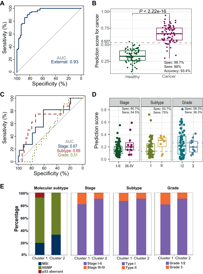
Results: The liquid biopsy assay demonstrated high efficacy in early EC detection, achieving an area under the curve (AUC) of 0.96, with 75.8% sensitivity and 96.8% specificity in the independent test cohort. Consistent sensitivities were observed across EC stages I-IV at 74.4%, 85.7%, 75%, and 75%, respectively. The assay moderately predicted clinicopathological features including stage (AUC = 0.72), histological subtypes (AUC = 0.73), and microsatellite instability status (AUC = 0.77). The model also effectively predicted recurrence-free survival, identifying high-risk patients [hazard ratio (HR) 8.6, P < 0.001]. Additionally, similarity network fusion stratified patients into high- and low-risk clusters, with high-risk individuals exhibiting a notably increased recurrence risk (HR 6.2, P = 0.049). Patients identified as high-risk by both methods exhibited an even greater risk (HR 10.1, P < 0.0001) for recurrence.

Conclusions: This DECIPHER-UCEC-2 study (Detecting Early Cancer by Inspecting ctDNA Features) demonstrates that by integrating cfDNA fragmentomics with machine learning, our liquid biopsy assay shows significant promise for EC's early detection, subtyping, and prognosis, potentially paving the way for enhanced patient outcomes.

Keywords: cell-free DNA; early diagnosis; endometrial cancer; fragmentomics; whole-genome sequencing.


Figures

Figure 1
Figure 1
Cohort description and flowchart. Workflow of early detection of endometrial cancer (A) and prediction of clinicopathological phenotypes of endometrial cancer based on preoperative cfDNA fragmentomics features (B). cfDNA, cell-free DNA; CNV, copy number variation; DL, deep learning; FSD, fragment size distribution; FSS, fragment size score; GBM, gradient boosting machine; GLM, generalized linear model; MCMS, mutation context and mutational signature; NF, nucleosome footprint; RF, random forest.
Figure 2
Figure 2
Early detection of endometrial cancer by stacking modeling based on integrated cfDNA fragmentomics features. (A) ROC curves of the training cohort and test cohort. The prediction score of each sample in the training cohort was the average score derived from the models with ≥0.6 of average AUC in the five-fold cross-validation repeating 10 times. The prediction score of each sample in the test cohort was the average score from the models with ≥0.6 of average AUC reconstructed based on the whole training cohort. (B) Difference of prediction scores between cancer patients and healthy subjects in the training cohort and test cohort. 0.527 is the point that maximizes the Youden index. AUC, area under the curve; cfDNA, cell-free DNA; ROC, receiver operating characteristic; Sens, sensitivity; Spec, specificity.
Figure 3
Figure 3
Early detection and prediction of endometrial cancer by integrated cfDNA fragmentomics features in the external cohort. (A) ROC curves of the external cohort. The prediction score of each sample in the external cohort was the average score from the models with ≥0.6 of average AUC reconstructed based on the whole training cohort. (B) Difference in prediction scores between cancer patients and healthy subjects in the external cohort. (C) External ROCs for the prediction of FIGO stage, histological subtypes, and differentiation grades by cfDNA fragmentomics features. (D) The distribution of prediction scores with FIGO stages, histological subtypes, and differentiation grades. (E) Distribution of FIGO stage, histological subtypes, and differentiation grades in the two clusters. AUC, area under the curve; cfDNA, cell-free DNA; FIGO, International Federation of Gynecology and Obstetrics; MSI, microsatellite instability; NSMP, no specific molecular profile; ROC, receiver operating characteristic; Sens, sensitivity; Spec, specificity.
Figure 4
Figure 4
Prediction of clinicopathological features of endometrial cancer by integrated cfDNA fragmentomics profiling. (A) Training and testing ROCs for the prediction of FIGO stage by cfDNA fragmentomics features. (B) The distribution of prediction scores with FIGO stages. 0.163 is the point maximizing the Youden index in the training cohort. (C) Training and testing ROCs for the prediction of histological subtypes by cfDNA fragmentomics features. (D) The distribution of prediction scores with histological subtypes. 0.139 is the point maximizing the Youden index in the training cohort. (E) Training and testing ROCs for the prediction of differentiation grade by cfDNA fragmentomics features. (F) The distribution of prediction scores with differentiation grades. 0.264 is the point maximizing the Youden index in the training cohort. (G) ROC for the prediction of MSI by cfDNA fragmentomics features through LOOCV. (H) The distribution of prediction scores with MSI status. 0.126 is the point maximizing the Youden index in the training cohort. AUC, area under the curve; cfDNA, cell-free DNA; FIGO, International Federation of Gynecology and Obstetrics; LOOCV, leave-one-out cross-validation; MSI, microsatellite instability; ROC, receiver operating characteristic; Sens, sensitivity; Spec, specificity; AIC, akaike information criterion.
Figure 5
Figure 5
Prediction of RFS in patients with endometrial cancer by integrated cfDNA fragmentomics profiling. (A) Estimation of patients’ recurrence-free survival by the Kaplan–Meier method in the training cohort. The patients were divided into high-risk group and low-risk group with the 67th percentile of the risk score (0.421) obtained from the LASSO Cox model as cut-off value. (B) Estimation of patients’ recurrence-free survival by the Kaplan–Meier method in the test cohort. The patients were divided into high-risk group and low-risk group with the same cut-off value as the training cohort. (C) Estimation of patients’ recurrence-free survival by the Kaplan–Meier method by including all patients. (D) Multivariate Cox regression analysis of recurrence-free survival. Risk score, HE4: human epididymis protein 4, CA125: carbohydrate antigen 125, and CA199: carbohydrate antigen 199 were treated as continuous variables and stage, grade, and subtype were treated as binary variables. AIC, akaike information criterion; cfDNA, cell-free DNA; EC, endometrial cancer; HR, hazard ratio; LASSO, least absolute shrinkage and selection operator; RFS, recurrence-free survival. ∗P < 0.05; ∗∗P < 0.01.
Figure 6
Figure 6
SNF clustering of endometrial cancer based on integrated cfDNA fragmentomics profiling. (A) Spectral clustering the endometrial cancer patients (n = 120) in the training cohort by SNF. The color in the cell represents the similarity between pairwise samples. (B) Distribution of grades in the two clusters. The clusters of the samples in the test cohort (n = 62) were predicted with a semi-supervised learning approach (label propagation), and the analysis includes all patients (n = 182) (same hereinafter). The Cochran–Armitage test was used for trend test (one-sided). (C) Distribution of histological subtypes in the two clusters. Fisher’s test was used to test the association between SNF cluster and subtype. (D) Distribution of FIGO stages in the two clusters. The Cochran–Armitage test was used for trend test (one-sided). (E) Multivariate logistic regression analysis of the association of SNF clusters with various clinicopathological features. SNF clusters were treated as the dependent variable and all patients were included. (F) The difference in the recurrence survival analysis between the two clusters estimated by the Kaplan–Meier method in the training cohort. (G) The difference in the recurrence survival analysis between the two clusters estimated by the Kaplan–Meier method in the test cohort. (H) The difference in the recurrence survival analysis between the two clusters estimated by the Kaplan–Meier method by including all patients. (I) Multivariate Cox regression analysis of recurrence-free survival. CA125, HE4, and CA199 were treated as continuous variables and SNF cluster, stage, grade, and subtype were treated as binary variables. (J) The patients were divided into four groups according to their SNF cluster and the group based on the risk score obtained from the LASSO Cox model. The recurrence-free survival of the four groups was estimated by the Kaplan–Meier method. AIC, akaike information criterion; cfDNA, cell-free DNA; EC, endometrial cancer; FIGO, International Federation of Gynecology and Obstetrics; LASSO, least absolute shrinkage and selection operator; SNF, similarity network fusion. ∗P < 0.05; ∗∗P < 0.01.

References

    1. Siegel R.L., Miller K.D., Fuchs H.E., Jemal A. Cancer statistics, 2022. CA Cancer J Clin. 2022;72(1):7–33. - PubMed
    1. Siegel R.L., Miller K.D., Jemal A. Cancer statistics, 2020. CA Cancer J Clin. 2020;70(1):7–30. - PubMed
    1. Oaknin A., Bosse T.J., Creutzberg C.L., et al. Endometrial cancer: ESMO Clinical Practice Guideline for diagnosis, treatment and follow-up. Ann Oncol. 2022;33(9):860–877. - PubMed
    1. Frias-Gomez J., Benavente Y., Ponce J., et al. Sensitivity of cervico-vaginal cytology in endometrial carcinoma: a systematic review and meta-analysis. Cancer Cytopathol. 2020;128(11):792–802. - PubMed
    1. Jacobs I., Gentry-Maharaj A., Burnell M., et al. Sensitivity of transvaginal ultrasound screening for endometrial cancer in postmenopausal women: a case-control study within the UKCTOCS cohort. Lancet Oncol. 2011;12(1):38–48. - PubMed

LinkOut - more resources