. 2025 Sep 24;16(1):8372.
doi: 10.1038/s41467-025-62405-6.

CD103-CD8+ T cells promote neurotoxic inflammation in Alzheimer's disease via granzyme K-PAR-1 signaling

Affiliations

CD103-CD8+ T cells promote neurotoxic inflammation in Alzheimer's disease via granzyme K-PAR-1 signaling

Eleonora Terrabuio et al. Nat Commun. .

Abstract

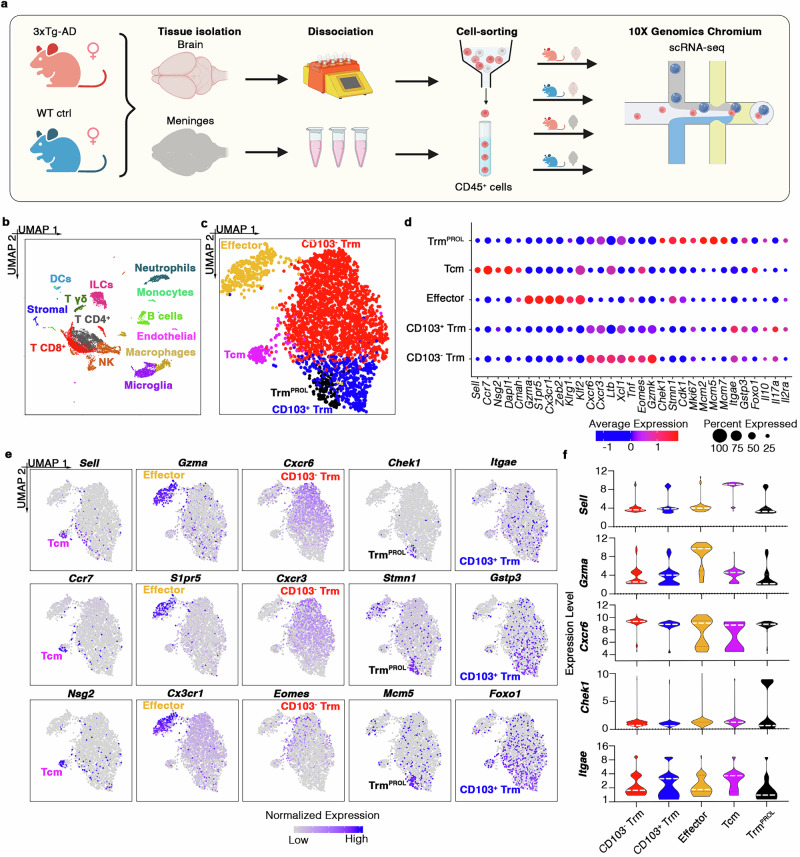
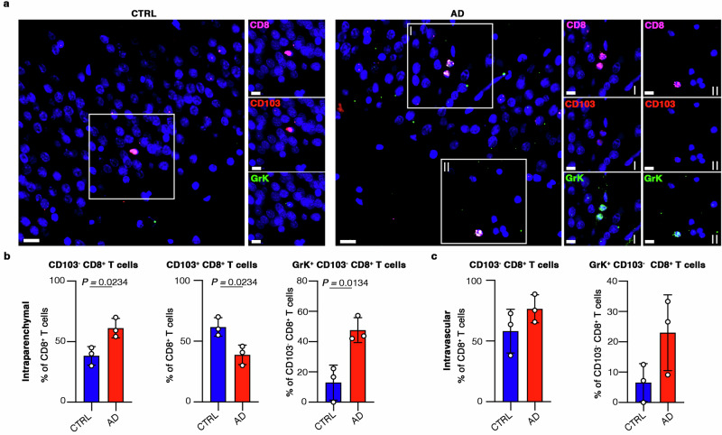
Immune mechanisms contribute to the neuropathology of Alzheimer's disease (AD) but the role of adaptive immune cells is unclear. Here we show that the brain CD8+ T cell compartment is dysregulated in AD patients and in the 3xTg-AD mouse model, accumulating activated CD103- tissue-resident memory T cells that produce large amounts of granzyme K (GrK). These CD103-CD8+ T cells originate from the circulation and migrate into the brain using LFA-1 integrin. Ablation of brain CD103-CD8+ T cells in 3xTg-AD mice ameliorates cognitive decline and reduces neuropathology. GrK induces neuronal dysfunction and tau hyperphosphorylation in human and mouse cells via protease-activated receptor-1 (PAR-1), which is expressed at higher levels in the AD brain, revealing a key immune-mediated neurotoxic axis. We conclude that communication between CD8+ T cells and the nervous system is altered in AD, paving the way for therapies targeting T cell-dependent neurotoxic inflammation.


Conflict of interest statement

Competing interests: The authors declare no competing interests.

Figures

Fig. 1
Fig. 1. Characterization of CD8 T cell phenotype in 3xTg-AD and WT mice.
a Graphical overview of the scRNAseq experimental design created in BioRender. Terrabuio, E. (2025) https://BioRender.com/f3lmkb2. b UMAP plot showing CD45HIGH leukocytes detected in the brains and meninges of wild-type (WT; n = 8) and 3xTg-AD (n = 8) mice. CD8+ T cells (n = 3,098) are shown in red. c UMAP plots showing the five subsets of CD8+ T cells detected in the brains and meninges of WT (n = 8) and 3xTg-AD (n = 8) mice. d Bubble plot reporting the phenotypic marker genes for each CD8+ T cell subset. Transcript levels are color-coded. e Normalized expression of known marker genes on UMAP plot. Transcript levels are color-coded. f Violin plots showing the expression of marker genes for each CD8+ T cell subset. White dashed line indicates the median expression level.
Fig. 2
Fig. 2. The CD8+ Trm compartment is altered in the brains of 3xTg-AD mice.
a, b Violin plots (g) and bubble plot (h) showing the expression of phenotypic markers detected by flow cytometry in the brains and meninges of WT (n = 3 pools of two organs each) and 3xTg-AD (n = 3 pools of two organs each) mice. White dashed line indicates the median expression level in violin plots (g). MFI expressions in the bubble plot (h) are scaled and color-coded. P-values are based on two-way ANOVA - multiple comparisons. Source data are provided as a Source Data file. c Donut plots indicating the distribution of CD8+ T cell subsets in the brains of WT (left) and 3xTg-AD (right) mice d Heat map showing the expression of residency genes in the brains and meninges of WT and 3xTg-AD mice. Transcript levels are color-coded. e Violin plots showing gene expression in the brains of WT (n = 8) and 3xTg-AD (n = 8) mice. f Heat map showing the expression in CD45HIGH leukocytes of genes in brains of WT (n = 8) and 3xTg- AD (n = 8) mice. Transcript levels are color-coded.
Fig. 3
Fig. 3. GrK expression is upregulated in brain CD103CD8+ Trm cells of 3xTg-AD mice.
a Pseudo-time ordered trajectory plot of CD8+ T cells in the brains and meninges of WT and 3xTg- AD mice. b Expression of genes in the trajectory plots. Transcript levels are color-coded. c Trajectory plot indicating the distribution of brain CD103 and CD103+CD8+ Trm cells in WT and 3xTg-AD mice. d, e Abundances of brain CD103 (d) and CD103+ (e) CD8+ Trm cells in WT (n = 4) and 3xTg-AD (n = 4) mice. Data are means ± SD. P-values based on two-tailed Student’s t-test. f Scatter plot showing KEGG pathway enrichment analysis for CD103 and CD103+ CD8+ Trm cells in the brains of 3xTg-AD mice (PD = “Parkinson disease”, ALS = “Amyotrophic lateral sclerosis”). g Bubble plot showing BP-GO GSEA analysis for brain CD103CD8+ Trm cells in 3xTg-AD mice. h Normalized gene expression on UMAP plot of brain and meningeal CD8+ T cells. Transcript levels are color-coded. i Violin plots showing gene expression in CD103 and CD103+CD8+ Trm cells in the brain and meninges. j Violin plot showing gene expression in CD8+ T cells in the brains of WT and 3xTg-AD mice. k Representative flow cytometry plot showing GrK expression in brain CD8+ T cells in WT and 3xTg-AD mice. l Bar plots showing the percentage of GrK, GrKMED, and GrKHIGH CD103 CD8+ Trm cells in the brains of WT (n = 5) and 3xTg-AD (n = 5) mice. Data are means ± SD. P-values based on two-tailed Student’s t-test. m Scatter plot showing MFIs of CD103 and GrK in CD8+ T cells in the brains of WT (n = 5) and 3xTg-AD (n = 5) mice. n, o Representative histograms (n) and violin plots (o) showing GrK MFI for brain CD103 (red) and CD103+ (blue) CD8+ Trm cells in 3xTg-AD mice. White dashed lines indicate median expressions. P-values based on two-tailed Student’s t-test. p CD8+ T cells (squares) in the brains of WT and 3xTg-AD mice detected by immunofluorescence microscopy. Scale bar = 5 µm or 2 µm for zoomed images.
Fig. 4
Fig. 4. GrK+CD103CD8+ Trm cells induce neuronal alterations by engaging PAR-1.
a Graphical overview of the experimental design. Created in BioRender. Terrabuio, E. (2025) https://BioRender.com/ir5h5rf. b Immunofluorescence staining showing GrK granules inside CD103+ and CD103 CD8+ Trm cells. Scale bar = 3 µm. c Intracellular Ca2+ release in neurons co-cultured with CD103+ or CD103 CD8+ Trm cells. The last time point is shown in the right panel. Data are means ± SD from four independent experiments. P-values based on two-way ANOVA-multiple comparisons. Ctrl = neurons alone. Ctrl+ = ionomycin-stimulated neurons (10 µM). Source data are provided as a Source Data file. d Intracellular Ca2+ release in neurons treated with purified active GrK or vehicle. The last time point is shown in the right panel. Data are means ± SD from three independent experiments. P-values based on two-way ANOVA-multiple comparisons. Source data are provided as a Source Data file. e Intracellular Ca2+ release in neurons co-cultured for 5 h with CD103CD8+ Trm cells in the presence/absence of the PAR-1 inhibitor SCH79797 (100 nM). The last time point (300 min) is shown in the right panel. Data are means ± SD from four independent experiments. P-values based on two-way ANOVA-multiple comparisons. Ctrl = neurons alone. Ctrl+ = ionomycin-stimulated neurons (10 µM). Source data are provided as a Source Data file. f, g Intracellular Ca2+ release in neurons cultured for 5 h with purified active GrK alone (150 nM) or with SCH79797 (50 nM, or 100 nM). Ctrl = neurons alone. The last time point is shown in the right panel. Data are means ± SD from three independent experiments. P-values based on two-way ANOVA-multiple comparisons. Representative images are shown in (g). Red = intracellular Ca2+ release. Scale bar = 20 µm. Source data are provided as a Source Data file. h Immunofluorescence microscopy showing GrK+CD103CD8+ Trm cells near the soma (cell I) and dendrites (cell II) of a PAR-1+ neuron. Cell morphology was visualized by wide-field imaging using a DIC filter. Scale bar = 5 µm (or 2 µm for zoomed images I, and II).
Fig. 5
Fig. 5. Detrimental CD103CD8+ Trm cells in the brain originate from the circulation and their depletion ameliorates disease in 3xTg-AD mice.
a Flow cytometry showing the percentage of brain CD103 and CD103+CD8+ Trm cells after anti-CD8 antibody treatment. Anti-CD8 (WT, n = 6; 3xTg-AD, n = 9), isotype-control (WT, n = 6; 3xTg-AD, n = 9). Data are means ± SD from two independent experiments and P-values are based on one-way ANOVA-multiple comparisons was used. b Time spent during training (MWM test). Anti-CD8 (WT, n = 10; 3xTg-AD, n = 21), isotype-control (WT, n = 22; 3xTg-AD, n = 18). Data are means ± SD from two-independent experiments (two-way ANOVA-multiple comparisons). c Representative tracking of three mice/group (MWM test). d, e Violin plots showing path efficiency (d) and number of body rotations (e) (MWM test). Data are from two-independent experiments one-way ANOVA-multiple comparisons). f Bar plot showing the percentage of freezing during the CFC test after anti-CD8 treatment. Anti-CD8 (WT, n = 9; 3xTg-AD, n = 16), isotype-control (WT, n = 21; 3xTg-AD, n = 13). Data are means ± SD from two-independent experiments. One-way ANOVA-multiple comparisons was used. gi Immunohistochemical staining of the hippocampus after CD8 T cell depletion showing Aβ-load (g), the levels of hyperphosphorylated (h) and total (i) tau protein. Anti-CD8 (n = 3), isotype-control (n = 3). Scale bar = 20 µm. Data are means ± SEM. Two-tailed Student’s t-test was used. ROI = 624.7 µm×501.22 µm. jl ELISA showing Aβ 1−40 and Aβ 1−42 (j) levels of tau hyperphosphorylation (k) and total tau (l) in soluble and insoluble fractions of brain homogenates after anti-CD8 treatment. Anti-CD8 (n = 4), isotype control (n = 4). Data from three-independent experiments are means ± SEM (two-tailed Student’s t-test). m Dot blot showing insoluble oligomeric and fibrillar forms of Aβ in brain homogenates after anti-CD8 treatment. Anti-CD8 (n = 4), isotype-control (n = 4). Data from three-independent experiments are shown as means ± SEM. P-values based on two-tailed Student’s t-test. n Immunofluorescence staining showing hippocampal oligomeric and fibrillar Aβ. Scale bar = 10 µm.
Fig. 6
Fig. 6. LFA-1 integrin controls the accumulation of CD8+CD103 cells in the brains of 3xTg- AD mice.
a Flow cytometry showing the percentage of LFA-1+ CD8+ T cells (left) and corresponding MFIs (right) in the blood of WT (n = 6) and 3xTg-AD (n = 6) mice. Data are means ± SD, with P-values based on two-tailed Student’s t-test. b Flow cytometry showing the percentage of CD45+ leukocytes among live cells. Data are means ± SEM, with P-values based on two-tailed Student’s t-test. c Flow cytometry showing the percentage of CD103 (left) and CD103+ (right) CD8+ Trm cells in the brains of 3xTg-AD (n = 6) and 3xTg-AD/Itgal/ (n = 6) mice. Data are means ± SEM, with P-values based on two-tailed Student’s t-test. df Representative immunohistochemical staining of the hippocampus in 3xTg-AD (n = 3) and 3xTg-AD/Itgal/ (n = 3) mice, showing the Aβ load (c), and the levels of hyperphosphorylated (d) and total (e) tau protein. Scale bar = 20 µm. Data are means ± SEM, with P-values based on two-tailed Student’s t-test. g UMAP plot showing immune cell populations in human blood (n = 3 AD patients and n = 2 negative controls, NCs) by scRNAseq. h Normalized expression of CD3G, CD3D and CD3E on a UMAP plot. Transcript levels are color- coded i CD3+ T cell cluster subsets: UMAP plot showing CD8+ and CD4+ T cell subpopulations in human blood (n = 3 AD patients and n = 2 NCs). j Normalized expression of CD8A, CD8B and CD4 on a UMAP plot. Transcript levels are color- coded. k CD8+ T cell cluster subsets: UMAP plot showing early active, Tem, and naïve-like CD8+ T cells in human blood (n = 3 AD patients and n = 2 NCs). l Radar plot showing AUCell score using known genes. m Heat map reporting the expression values (mean calculated using the Hurdle model) of ITGAL in AD patients (n = 3) and NCs (n = 2). Transcript levels are color-coded.
Fig. 7
Fig. 7. GrK+CD103CD8+ Trm cells accumulate in the CSF of AD patients.
a UMAP plot showing leukocyte populations detected in human CSF (n = 4 AD, n = 5 MCI, and n = 9 healthy controls, HCs) by scRNAseq. b Normalized expression of CD3E, CD8B and CD4 in the UMAP plot. Transcript levels are color-coded. c CD8+ T cell cluster subsets: UMAP plot showing KLF2+ and Trm cell subpopulations in human CSF (n = 4 AD, n = 5 MCI, and n = 9 HCs). d Normalized expression of KLF2, CD69 and PRDM1 in a UMAP plot. Transcript levels are color-coded. e Trm cell cluster subsets: UMAP plot showing CD103 and CD103+ Trm cells in human CSF (n = 4 AD, n = 5 MCI, and n = 9 HCs). f Percentage of CD103 and CD103+ CD8+ cells in human CSF (n = 4 AD, n = 5 MCI, and n = 9 HCs). Data are means ± SD. Source data are provided as a Source Data file. g Violin plot reporting GZMK expression in Trm cells of n = 4 AD patients, n = 5 MCI subjects, and n = 9 HCs. The white dot represents median expression. h Normalized expression of GZMK in a UMAP plot of all Trm cells (panel 1) and Trm cells from HCs (panel 2), MCI subjects (panel 3), and AD patients (panel 4).
Fig. 8
Fig. 8. GrK+CD103CD8+ Trm cells accumulate in the brains of AD patients.
a Immunofluorescence staining of control (CTRL) and AD human brain tissues. Scale bar = 40 µm, or 15 µm in the zoomed images. b Bar plots showing the percentage of intraparenchymal CD103, CD103+, and GrK+CD103 CD8+ T cells in the hippocampus of HCs (n = 3) and AD patients (n = 3). Data are means ± SD (two-tailed Student’s t-test). Source data are provided as a Source Data file. c Bar plots showing the percentage of intravascular CD103and GrK+CD103 CD8+ T cells in the hippocampus of HCs (n = 3) and AD patients (n = 3). Data are means ± SD (two-tailed Student’s t-test). Source data are provided as a Source Data file.
Fig. 9
Fig. 9. GrK induces functional alterations in human neuronal cells via PAR-1.
a Immunofluorescence staining for βIII-tubulin, MAP-2, pNF-H, Nestin and PAR-1 in undifferentiated and differentiated SH-SY5Y human neuroblastoma cells. b Intracellular Ca2+ release in differentiated SH-SY5Y cells cultured in the presence of 150 nM GrK, or 150 nM GrK and 100 nM SCH79797. The last time point is shown in the bottom panel. Ctrl = neurons alone. Ctrl+ = ionomycin-stimulated neurons (10 µM). Data are means ± SEM from three independent experiments. P-values based on two-way ANOVA-multiple comparisons. c Representative images of differentiated SH-SY5Y cells at different time points cultured without GrK (Ctrl), in the presence of 150 nM active GrK, or in the presence of 150 nM active GrK plus 100 nM SCH79797. The red signal shows intracellular Ca2+ release. Scale bar = 20 µm.
Fig. 10
Fig. 10. GrK induces tau hyperphosphorylation and alteration of signaling pathways in human neuronal cells via PAR-1.
a–c Bar plots and representative images showing tau phosphorylation levels on serine and threonine (AT8, AT100) (d, e) and only threonine (AT180) (f) residues in differentiated SH-SY5Y cells cultured alone (Ctrl), in the presence of 100 nM SCH79797, in the presence of 150 nM active GrK alone, and the presence of both. Data are means ± SD of three independent experiments (two-way ANOVA-multiple comparisons). Scale bar = 5 µm. d ELISA showing the levels of phosphorylation on the pS199, pS396, and pT231 residues of tau protein in differentiated SH-SY5Y cells cultured alone (Ctrl), in the presence of 100 nM SCH79797, in the presence of 150 nM active GrK alone, and in the presence of both. Data are means ± SD of three independent experiments. P-values are based on two-way ANOVA multiple comparisons. e, f Protein enrichment analysis from three independent proteomic experiments showing the best enriched clusters of terms (h) and terms belonging to the “Alzheimer’s/neurodegeneration” and “kinase” pathways (i) in SH-SY5Y human neuroblastoma cells cultured in the absence of GrK and SCH79797 (Ctrl), in the presence of 100 nM SCH79797 alone, in the presence of 150 nM active GrK alone, and in the presence of both. Source data are provided as a Source Data file.

References

    1. Gustavsson, A. et al. Global estimates on the number of persons across the Alzheimer’s disease continuum. Alzheimers Dement19, 658–670 (2023). - PubMed
    1. Bettcher, B. M., Tansey, M. G., Dorothee, G. & Heneka, M. T. Peripheral and central immune system crosstalk in Alzheimer disease - a research prospectus. Nat. Rev. Neurol.17, 689–701 (2021). - PMC - PubMed
    1. Mary, A., Mancuso, R. & Heneka, M. T. Immune activation in alzheimer disease. Annu Rev. Immunol.42, 585–613 (2024). - PubMed
    1. Kaech, S. M. & Cui, W. Transcriptional control of effector and memory CD8+ T cell differentiation. Nat. Rev. Immunol.12, 749–761 (2012). - PMC - PubMed
    1. Park, S. L., Gebhardt, T. & Mackay, L. K. Tissue-Resident Memory T Cells in Cancer Immunosurveillance. Trends Immunol.40, 735–747 (2019). - PubMed

MeSH terms