. 2025 Sep 24;16(1):8363.
doi: 10.1038/s41467-025-62285-w.

Mechanically activated snai1b coordinates the initiation of myocardial delamination for trabeculation

Affiliations

Mechanically activated snai1b coordinates the initiation of myocardial delamination for trabeculation

Jing Wang et al. Nat Commun. .

Abstract

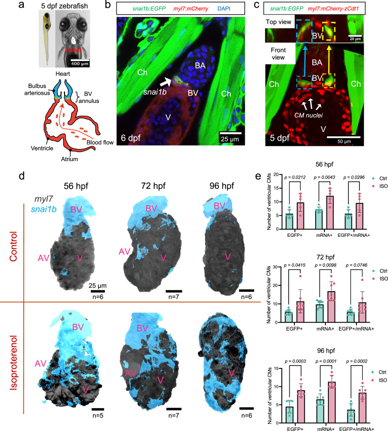
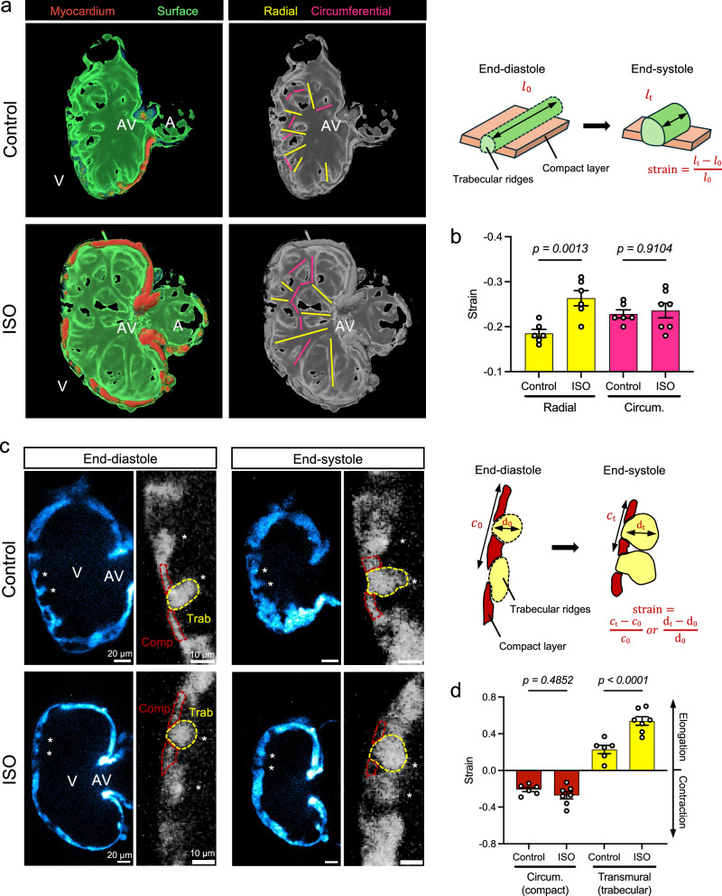
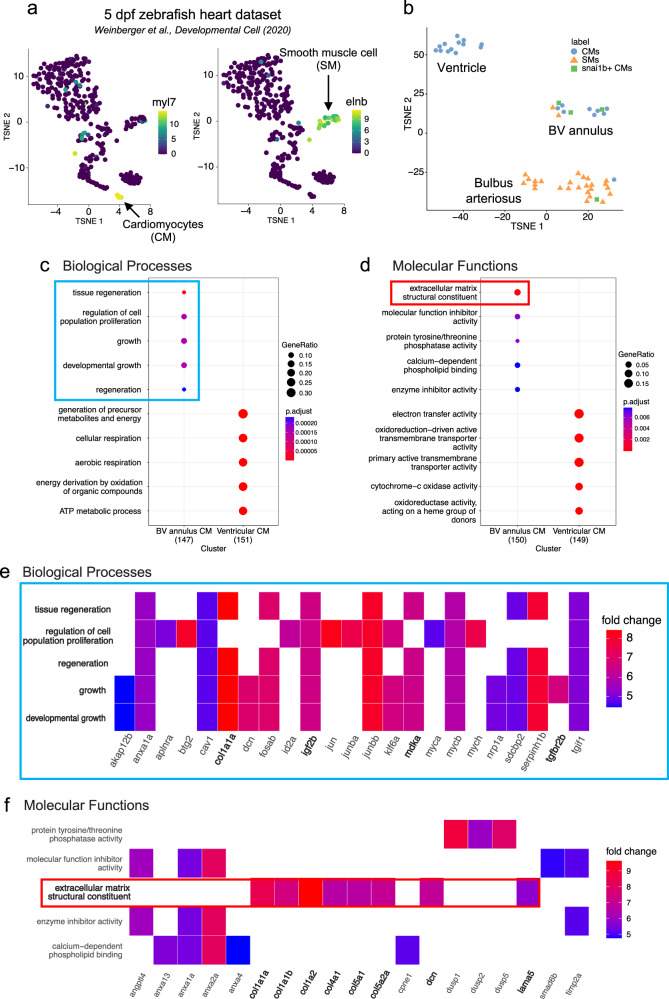
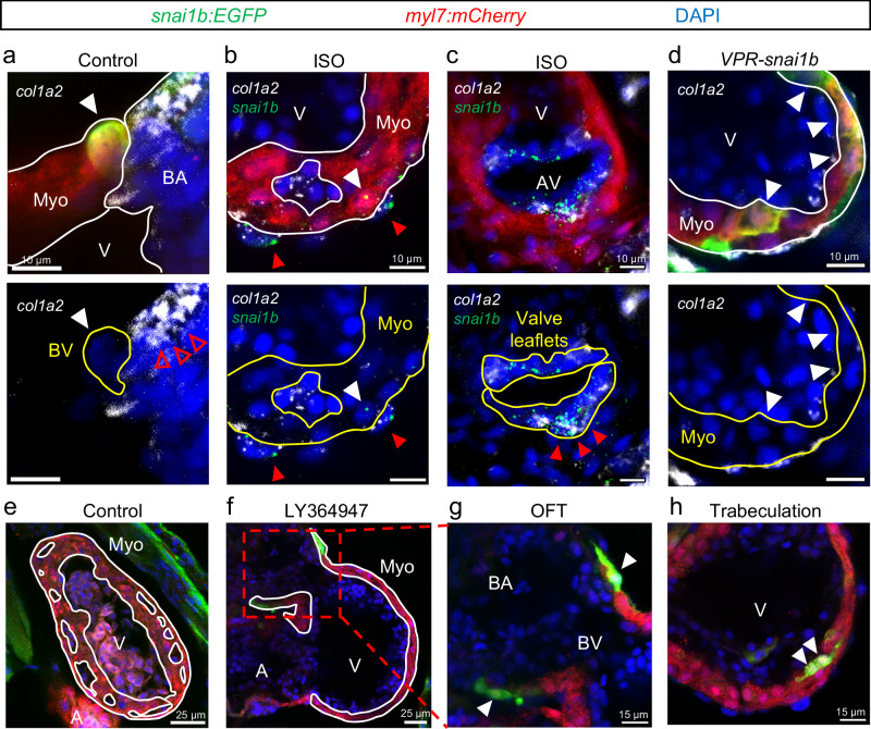
During development, myocardial contractile force and intracardiac hemodynamic shear stress coordinate the initiation of trabeculation. While Snail family genes are well-recognized transcription factors of epithelial-to-mesenchymal transition, snai1b-positive cardiomyocytes are sparsely distributed in the ventricle of zebrafish at 4 days post-fertilization. Isoproterenol treatment significantly increases the number of snai1b-positive cardiomyocytes, of which 80% are Notch-negative. CRISPR-activation of snai1b leads to 51.6% cardiomyocytes forming trabeculae, whereas CRISPR-repression reduces trabecular cardiomyocytes to 6.7% under isoproterenol. In addition, 36.7% of snai1b-repressed cardiomyocytes undergo apical delamination. 4-D strain analysis demonstrates that isoproterenol increases the myocardial strain along radial trabecular ridges in alignment with the snai1b expression and Notch-ErbB2-mediated trabeculation. Single-cell and spatial transcriptomics reveal that these snai1b-positive cardiomyocytes are devoid of some epithelial-to-mesenchymal transition-related phenotypes, such as Col1a2 production and induction by ErbB2 or TGF-β. Thus, we uncover snai1b-positive cardiomyocytes that are mechanically activated to initiate delamination for cardiac trabeculation.


Conflict of interest statement

Competing interests: The authors declare no competing interests.

Figures

Fig. 1
Fig. 1. Myocardial snai1b expression at the BV annulus and in the ventricle during trabeculation.
a A zebrafish embryo (left panel) at 5 dpf, and its heart is highlighted. Right panel illustrates the two-chambered cardiac anatomy. b At 6 dpf, confocal imaging of hearts of Tg(snai1b:EGFP; myl7:mCherry) larvae. The intensity of snai1b expression (green, arrow) was highest at the BV annulus (n = 5). c At 5 dpf, confocal imaging of hearts from Tg(myl7:mCherry-zCdt1) embryos (n = 7) demonstrates that snai1b fluorescence (colored squares) was colocalized to CM nuclei. Upper panel provides a view of the BV annulus from the BA. d During trabeculation (56–96 hpf), snai1b:EGFP signal is concentrated around the BV annulus. Isoproterenol (ISO) treatment at 1 dpf induced snai1b activation in the ventricular CMs. e Whole-mount in situ hybridization of snai1b mRNA in the Tg(snai1b:EGFP; myl7:mCherry) reporter line reveals a significant increase of both EGFP+ and mRNA+ CMs in ISO-treated hearts. (see Supplementary Figs. 1 and 2 for staining images). All values are displayed with mean and standard deviation (SD). p-value is displayed for each comparison. The number of hearts analyzed in each is displayed in (d). Ordinary two-way ANOVA followed by Šídák’s multiple comparisons test on the means was applied to determine statistical significance. Source data are provided as a Source Data file. Anatomic labels: BA bulbus arteriosus, V ventricle, BV bulbus-ventricular annulus, Ch chest wall, AV atrioventricular canal.
Fig. 2
Fig. 2. ISO-mediated increase in ventricular contractility and strain activates myocardial snai1b.
a, b At 14 dpf, the expression of myocardial snai1b (dashed green outline) remained sparse in the ventricle, whereas Isoproterenol (ISO) treatment from 1 to 11 dpf revealed a persistent snai1b activation in the trabecular network. Anatomic labels: BA bulbus arteriosus, V ventricle, BV bulbus-ventricular annulus, Myo myocardium, Endo endocardium, Epi epicardium, Ery erythrocyte. c At 4 dpf, 4-D mapping of myocardial strain in a control and an ISO-treated heart. Two time points during diastole were displayed, and the ISO-treated heart experienced higher strain at the end-diastole time point than the control. Red dashed lines and squares mark the cross-section planes, and the red arrows indicate the viewing direction. Blue and green arrows indicate the direction of blood flow. Anatomic labels: V ventricle, AV atrioventricular canal, OFT outflow tract. d Average epicardial/endocardial ventricular strain within the sampling regions during one cardiac cycle. ISO treatment increased the myocardial strain across the endocardial surface during the end-diastolic and systolic phases. Source data are provided as a Source Data file. e Strain variations within the sampling regions, calculated as the standard deviation of strains at each time point (phase). ISO treatment induced greater myocardial strain variations compared with the control heart. Source data are provided as a Source Data file.
Fig. 3
Fig. 3. ISO-mediated increase in strain aligning with radial trabecular ridges.
a, b 3-D volumetric and surface rendering revealed the network of radial (yellow) and circumferential (pink) trabecular ridges inside the hearts at 4 dpf. ISO treatment significantly increased the strain (i.e., shortening) along the radial trabecular ridges (n = 6 ridges for each group) but not the circumferential ridges (n = 6 ridges for control, n = 7 for ISO). c, d 2-D cross-sections of the trabeculae (asterisks) revealed that the surrounding compact myocardium (red dashed outlines) contracts circumferentially while the ridges (yellow dashed outlines) thicken (elongate) transmurally during systole. ISO treatment significantly increased the transmural strain within trabecular ridges (n = 6 ridges for control, n = 7 for ISO), whereas the circumferential strain in the surrounding compact layer remained similar (n = 6 ridges for control, n = 7 for ISO). Anatomic labels: V ventricle, A atrium, AV AV canal. All values are displayed with mean and standard error of mean (SEM). p-value is displayed for each comparison. One control heart and one ISO-treated heart were used for analysis. Ordinary one-way ANOVA followed by Šídák’s multiple comparisons test on the means was applied to determine statistical significance. Source data are provided as a Source Data file.
Fig. 4
Fig. 4. snai1b activation in Notch-negative cardiomyocytes undergoing delamination and trabeculation.
a At 4 dpf, myocardial Notch signaling (TP1) develops at the outer curvature of compact (cortical) layer, and discontinuation was observed when trabeculation sprouting occurs (dashed outlines in magnified sub-panels). Endocardial Notch mediates the ErbB2 signaling in trabecular cardiomyocytes (CMs) to activate Notch in the adjacent CMs and inhibit their delamination. On average, 1.3 snai1b+ CMs were found per ventricle (see (e), n = 6). b After ISO treatment, the gap between Notch-positive (+) CMs widened, and snai1b activation (arrowheads, via in situ hybridization) was observed in the Notch-negative () CMs in the compact layer (29.5%) and trabeculation (50%), and a small number of apically delaminated CMs (3.4%) (see Fig. S3a–c). On average, 12.6 snai1b+ CMs were found per ventricle (see e, f, n = 7). c Co-treatment with ErbB2 inhibitor, PD168393, reduced trabeculation (see Fig. S3d) but did not inhibit snai1b activation (arrowheads, see e, f). d Schematic summarizing the Notch and snai1b expression pattern in (ac). e, f At 4 dpf, ISO treatment led to a 10-fold increase in the total number of snai1b+ CMs per ventricle. ErbB2 inhibitor (PD) co-treatments at 55 hpf (n = 5) or 72 hpf (n = 7) did not change the total number of snai1b+ CMs. Rather, they reduced the total trabecular population from 50 to 37% and 39%, respectively. All values in panel e are displayed with mean and standard error of mean (SEM). p-value is displayed for each comparison. Ordinary one-way ANOVA followed by Šídák’s multiple comparisons test on the means was applied to determine statistical significance. Source data are provided as a Source Data file. Anatomic labels: V ventricle, A atrium, AV atrioventricular canal.
Fig. 5
Fig. 5. Activation and repression of snai1b modulates delamination for trabeculation.
a Experimental plan for the myocardial Tol2-CRISPR activation/interference (CRISPRa/i) system. Tol2 transposon plasmids were injected in one-cell stage embryos to induce mosaic activation or repression of snai1b among cardiomyocytes (CMs). In total, four sgRNAs that target different promoter regions of snai1b were used. Two scramble sgRNAs were used as control. Right panels show the mechanism of activation (VPR) and repression (KRAB). For system validation, see Supplementary Fig. 8. Partially created in BioRender. Wang, J. (2025) https://BioRender.com/ep5rh7k. bd Under ISO treatment, the majority of control (58.3%) and snai1b-repressed (56.7%) CMs remained in the compact layer at 4 dpf (96 hpf). Activation of snai1b led to significantly more delaminated (see Supplementary Fig. 9b) and trabecular CMs per heart (c), where 51.6% of total snai1b-activated CMs form trabeculae, compared to 25.0% of control and 6.7% of snai1b-repressed CMs (d). On the other hand, repression of snai1b resulted in 36.7% of CMs undergoing apical delamination. For representative images of CRISPRa/i-injected hearts without ISO, see Supplementary Fig. 9a. All values in (c) are displayed with mean and standard error of mean (SEM). p-value is displayed for each comparison. Number of hearts analyzed: Control-ISO = 7, Repression-ISO = 7, Activation-ISO = 11. Ordinary one-way ANOVA followed by Holm-Šídák’s multiple comparisons test on the means was applied to determine statistical significance. Source data are provided as a Source Data file. Anatomic labels: V ventricle, Comp compact layer, Trab trabeculae.
Fig. 6
Fig. 6. Enrichment of mesenchymal genes in snai1b+ CMs.
a, b The CMs and SMs were isolated from a single-cell RNA seq dataset of 5-dpf hearts. These cells produced a sub-dataset with three clusters. snai1b+ CMs (green squares) were found in a cluster with a mixture of CMs and SMs, resembling the BV annulus region. The color scale indicates the normalized log counts of genes. The number of cells in the complete dataset was 366 after filtering, and the sub-dataset contains 53 cells. c, d Gene Ontology (GO) enrichment analysis categorized the enriched Biological Processes (BP) and Molecular Functions (MF). The BV annulus CMs had enriched mesenchymal BPs and MFs (blue and red boxes), compared to the ventricular CMs. The total number of marker genes selected for GO analysis was 151 for Ventricular CM and 147 for BV annulus CMs. The dot size indicates the gene ratio (i.e., # of genes in a category divided by # of total genes). The color scale depicts the Benjamini & Hochberg (1995) adjusted p-values. The original p-values are calculated using a hypergeometric test (one-tailed) via the enrichGO function in clusterProfiler. Top five enriched terms with p-values < 0.05 from each group are displayed. Source data are provided as a Source Data file. e, f Heatmaps showing the genes involved in each of the BP and MF categories for BV annulus CMs. The color scale indicates the log-fold change in mean expression for each gene in BV annulus CMs compared to ventricular CMs. Top five enriched terms with p-values < 0.05 are displayed. Source data are provided as a Source Data file.
Fig. 7
Fig. 7. snai1b pathways in CMs vs. epicardial and valvular cells.
ad Abundant col1a2 mRNA colocalized with snai1b mRNA in epicardial, valvular cells (red arrowheads), and bulbus smooth muscle cells (empty red arrowheads). However, col1a2 expression was not observed in snai1b+ CMs (white arrowheads) of control (a), ISO-treated (b), and CRISPRa-injected (d) hearts at 4 dpf. Number of hearts: Control = 10, ISO = 6, CRISPRa (VPR) = 10. eh At 3 dpf, LY364947, a TGF-β signaling inhibitor, attenuated trabeculation (white outlines), but the expression of snai1b (arrowheads) within BV annulus and trabecular CMs persisted. Number of hearts: Control n = 10, LY364947 n = 12. BA bulbus arteriosus, V ventricle, BV bulbus-ventricular annulus, A atrium, Ch chest wall, Myo myocardium, AV atrioventricular canal.

References

    1. Samsa, L. A. et al. Cardiac contraction activates endocardial Notch signaling to modulate chamber maturation in zebrafish. Development142, 4080–4091 (2015). - PMC - PubMed
    1. Lee, J. et al. Spatial and temporal variations in hemodynamic forces initiate cardiac trabeculation. JCI Insight3, e96672 (2018). - PMC - PubMed
    1. Hsu, J. J. et al. Contractile and hemodynamic forces coordinate Notch1b-mediated outflow tract valve formation. JCI Insight4, e124460 (2019). - PMC - PubMed
    1. Priya, R. et al. Tension heterogeneity directs form and fate to pattern the myocardial wall. Nature588, 130–134 (2020). - PubMed
    1. Wang, M. et al. Shear and hydrostatic stress regulate fetal heart valve remodeling through YAP-mediated mechanotransduction. eLife12, e83209 (2023). - PMC - PubMed

Substances

LinkOut - more resources