[Preprint]. 2025 Apr 6:2025.04.06.647428.
doi: 10.1101/2025.04.06.647428.

The functional organization of chromosome territories in single nuclei during zygotic genome activation

Affiliations

The functional organization of chromosome territories in single nuclei during zygotic genome activation

Akshada Shankar Ganesh et al. bioRxiv. .

Update in

Abstract

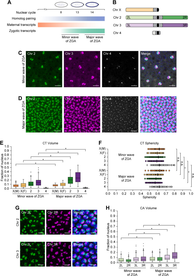
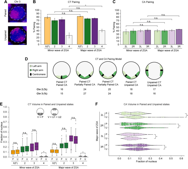
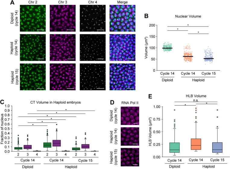
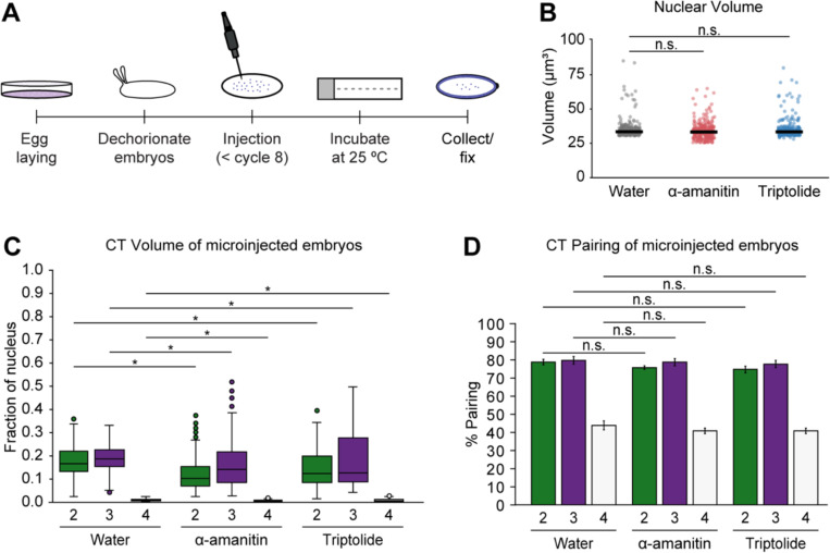
Chromosome territories (CTs) are intricately organized and regulated within the nucleus. Despite remarkable advances in our understanding of genome packaging and gene expression, the interplay among CTs, pairing of parental homologous chromosomes, and genome function during development remains elusive. Here, we employ an Oligopaints-based high-resolution imaging approach to examine variable CT organization in single nuclei during the developmental process of zygotic genome activation. We reveal large-scale chromosome changes with extensive homolog pairing at the whole-chromosome level that decreases locally due to spatial variability in chromosome conformations. In the absence of one homolog copy, the dynamics of CT compaction and RNA polymerase II recruitment are supported by transcriptional changes in haploid embryos. Finally, global inhibition of transcription results in decreased CT opening and no significant impact on CT pairing levels. These findings enhance our understanding of parental genome folding and regulation, which may inform strategies for chromosome-based diseases.

Keywords: RNA polymerase II; chromosome territories (CTs); haploid; homolog pairing; transcription; zygotic genome activation (ZGA).


Conflict of interest statement

Declaration of Interests The authors declare no competing interests.

Figures

Figure 1.
Figure 1.. Changes in CTs and CAs during the minor and major waves of ZGA
(A) Schematic representation of events during the onset of minor (nuclear cycle 8) and major (nuclear cycle 14) waves of ZGA. Maternal transcripts (orange) decrease, while zygotic transcripts (green) and somatic homolog pairing (blue) progressively increase. (B) Oligopaint probes target Drosophila chromosomes X, 2, 3, and 4 along with the arms of major chromosomes labelled. Centromere, black; heterochromatin, dark gray. (C and D) Chromosomes 2 (green), 3 (magenta), and 4 (gray) of Drosophila embryos during the minor (C) and major (D) waves of ZGA. Total DNA by Hoechst stain (blue). Bar = 10 μm. (E) CT volume from the minor to major waves of ZGA. Each CT volume is normalized to the respective nuclear volume. X(M), chromosome X in males; X(F), chromosome X in females; at least three replicates; n ≥ 300 nuclei; *p ≤ 1.27×10⁻⁶, Mann-Whitney two-sided U test. (F) CT sphericity between the minor and major waves of ZGA. X(M), chromosome X in males; X(F), chromosome X in females; median, solid line; at least three replicates; n ≥ 300 nuclei; *p ≤ 7.33×10⁻¹³, n.s., not significant, Mann-Whitney two-sided U test. (G) Representative images display the left (green) and right (magenta) arms of chromosomes 2 (top) and 3 (bottom) in the major wave of ZGA. Total DNA by Hoechst stain (blue). Bar = 5 μm. (H) CA volume from the minor to major waves of ZGA. Each CA volume is normalized to the respective nuclear volume. At least three replicates; n ≥ 300 nuclei; *p ≤ 1.94×10⁻¹⁰, Mann-Whitney two-sided U test.
Figure 2.
Figure 2.. Extensive homolog pairing of CTs and CAs during ZGA
(A) Paired (top) and unpaired (bottom) chromosome 3 (magenta) during major wave of ZGA. Total DNA by Hoechst stain (blue). Bar = 1 μm. Percentage of nuclei showing (B) CT pairing and (C) CA pairing from the minor to major waves of ZGA. X(F), chromosome X in females; error bars, standard deviation; at least three replicates; n ≥ 300 nuclei; *p = 6.26×10⁻³, n.s., not significant, Fisher’s two-tailed exact test. (D) Schematic representation illustrating the percentage of occurrence in major wave of ZGA for the different conformations of left (light green) and right (dark green) arm, indicating higher CT than CA pairing. The angles between CAs may vary. (E) Normalized CT volume between paired homologs to the combined volume of two unpaired homologs from the minor and major waves of ZGA. X(F), chromosome X in females; P, paired; U, unpaired; U1, unpaired homolog 1; U2, unpaired homolog 2; V, volume; at least three replicates; n ≥ 88 nuclei; *p ≤ 8.89×10⁻⁷, n.s., not significant, Mann-Whitney two-sided U test. (F) Normalized CA volume differences in arms of paired homologs to the combined volume of two unpaired homologs during the major wave of ZGA. P, paired; U, unpaired; local minima, dashed line; at least three replicates; n ≥ 242 nuclei; *p ≤ 3.06×10⁻⁵, n.s., not significant, Levene’s test.
Figure 3.
Figure 3.. CT dynamics in haploid embryos during ZGA
(A) Chromosomes 2, 3, and 4 in diploid (cycle 14, top) and haploid embryos (cycle 14, middle; cycle 15, bottom) during major wave of ZGA. Total DNA by Hoechst stain (blue). Bar = 10 μm. (B) Nuclear volume between diploid (cycle 14) and haploid embryos (cycles 14 and 15). Median, solid line; at least three replicates; n ≥ 300 nuclei; *p ≤ 2.09×10⁻¹⁰, Mann-Whitney two-sided U test. (C) CT volume differences for chromosomes 2, 3, and 4 in diploid and haploid embryos. In diploid embryos, only individual unpaired homolog volumes were used. Each CT volume is normalized to the respective nuclear volume. At least three replicates; n ≥ 300 nuclei; *p ≤ 1.81×10⁻², Mann-Whitney two-sided U test. (D) RNA Pol II in diploid (cycle 14, top) and haploid embryos (cycle 14, middle; cycle 15, bottom) during major wave of ZGA. Bar = 5 μm. (E) HLB volume between diploid and haploid embryos. In diploid embryos, only individual unpaired homolog volumes were used. Median, solid line; at least three replicates; n ≥ 300 nuclei; *p ≤ 3.20×10⁻¹⁵, n.s., not significant, Mann-Whitney two-sided U test.
Figure 4.
Figure 4.. CTs and homolog pairing in transcription inhibited embryos
(A) Diagram illustrating embryonic microinjections. (B) Nuclear volume in control, alpha-amanitin, and triptolide-treated embryos. Median, solid line; at least three replicates; n ≥ 300 nuclei; n.s., not significant, Mann-Whitney two-sided U test. (C) Normalized CT volume for chromosomes 2, 3, and 4 in water (control), alpha-amanitin, and triptolide-treated embryos. At least three replicates; n ≥ 300 nuclei; *p ≤ 6.11×10⁻³, Mann-Whitney two-sided U test. (D) CT pairing of transcription inhibited and control embryos. Error bars, standard deviation; at least three replicates; n ≥ 300 nuclei; n.s., not significant, Fisher’s two-tailed exact test.

References

    1. Bickmore W.A., and van Steensel B. (2013). Genome architecture: domain organization of interphase chromosomes. Cell 152, 1270–1284. 10.1016/j.cell.2013.02.001. - DOI - PubMed
    1. Bolzer A., Kreth G., Solovei I., Koehler D., Saracoglu K., Fauth C., Muller S., Eils R., Cremer C., Speicher M.R., and Cremer T. (2005). Three-dimensional maps of all chromosomes in human male fibroblast nuclei and prometaphase rosettes. PLoS Biol 3, e157. 10.1371/journal.pbio.0030157. - DOI - PMC - PubMed
    1. Cremer T., and Cremer C. (2001). Chromosome territories, nuclear architecture and gene regulation in mammalian cells. Nat Rev Genet 2, 292–301. 10.1038/35066075. - DOI - PubMed
    1. Cremer T., and Cremer M. (2010). Chromosome territories. Cold Spring Harb Perspect Biol 2, a003889. 10.1101/cshperspect.a003889. - DOI - PMC - PubMed
    1. Erceg J., AlHaj Abed J., Goloborodko A., Lajoie B.R., Fudenberg G., Abdennur N., Imakaev M., McCole R.B., Nguyen S.C., Saylor W., et al. (2019). The genome-wide multi-layered architecture of chromosome pairing in early Drosophila embryos. Nat Commun 10, 4486. 10.1038/s41467-019-12211-8. - DOI - PMC - PubMed

Publication types