. 2025 Aug 19;28(9):113377.
doi: 10.1016/j.isci.2025.113377. eCollection 2025 Sep 19.

A magnetotactic bacterium capable of magnetic sensing

Affiliations

A magnetotactic bacterium capable of magnetic sensing

Emilie Gachon et al. iScience. .

Abstract

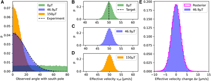
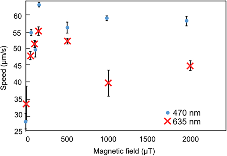
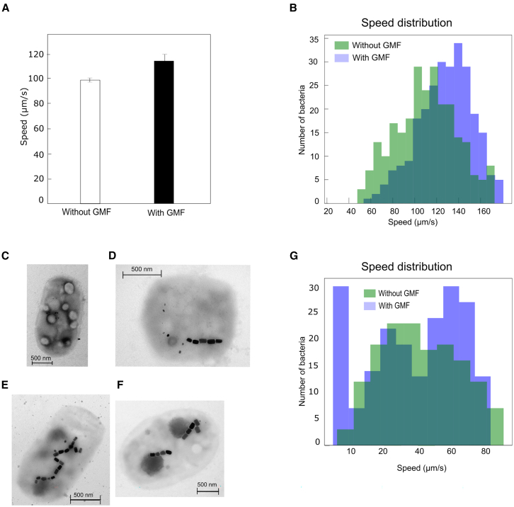
Magnetosensitive organisms have the ability to sense and respond actively to features of magnetic fields such as the direction or magnitude. Until now, magnetosensing has been characterized primarily in higher organisms, involving either a cryptochrome-based mechanism or direct magnetic interactions with magnetic particles. Magnetotactic bacteria, microorganisms forming intracellular chains of magnetic nanoparticles, are thought to only passively orient along field lines. In this study, we reveal that the cultivated magnetotactic bacterium SS-5 also exhibits magnetosensing. The microorganisms indeed swim faster in a physiological magnetic field compared to when the field is canceled. This speed difference is independent of illumination wavelength but is altered when the bacterial magnetic backbone is disrupted. We thus propose that magnetosensing in the bacteria originates from a magnetomechanical signal transduction along the magnetotactic filament. Our findings also show that this response depends on relative changes in magnetic field intensity, akin to the Weber-Fechner laws, suggesting that magnetosensing operates similarly to other forms of taxes.

Keywords: Evolutionary biology; Microbiology.


Conflict of interest statement

The authors declare no competing interests.

Figures

None
Graphical abstract
Figure 1
Figure 1
Trajectories of SS-5 bacteria in the presence and absence of the Earth’s magnetic field (A) 2D trajectories of individual cells in the presence and absence of a GMF. (B) Angular distribution of the direction of the trajectories with respect to the South magnetic pole.
Figure 2
Figure 2
SS-5 swimming speed as a function of magnetic field intensity and oxygen concentration (A) The average 3D speed of SS-5 bacteria imaged under a 585 nm illumination and measured in low oxygen concentrations (in media flushed with nitrogen for 10 min prior to imaging) and ambient oxygen concentrations increases in the presence of the Earth’s magnetic field and with increasing fields up to 150 μT before decreasing and plateauing for fields from 500 μT up to 3 mT. At least 50 cells were tracked per data point, and the error bars correspond to the standard error of the average speed of all the analyzed cells’ trajectories. From 0 mT to 3 mT, the average speed of bacteria swimming in ambient oxygen concentrations of oxygen are 38.7 ± 1.0 μm/s, 56.4 ± 0.4 μm/s, 53.1 ± 1.0 μm/s, 61.1 ± 2.9 μm/s, 47.8 ± 1.3 μm/s, 55.4 ± 0.7 μm/s, 48.1 ± 2.7 μm/s and 25.9 ± 2.0 μm/s, 30.9 ± 2.0 μm/s, 33.9 ± 2.0 μm/s, 32.4 ± 2.0 μm/s, 30.5 ± 1.0 μm/s, 28.9 ± 1.5 μm/s, and 30.9 ± 1.8 μm/s for bacteria in low oxygen concentrations. (B) Effect of different magnetic field intensities on SS-5’s bacterial flagellar motor’s rotational speed. Speeds were normalized with respect to the speed of the average speed of the bacterial flagellar motor in the absence of a GMF (without a GMF: 1.43 ± 0.2; 1 mT: 1.71 ± 0.1). Student’s t tests revealed no statistical differences for the bacterial flagellar motor’s rotational speed in the presence and absence of a GMF (p value of 0.39), whereas a p value of 0.003 was obtained when comparing the speed of the bacterial flagellar motors in the absence of a GMF and with an external magnetic field of 1 mT. A p value of 0.02 was obtained when comparing the rotation of the bacterial flagellar motor in the presence of a GMF and a field 1 mT.
Figure 3
Figure 3
Numerical study on the effect of a magnetic field on the effective swimming speed of a simulated swimmer undergoing helical movement (A) Distribution of the angle between the trajectory segments and the magnetic field lines. Depicted are the results for parameters that maximize the likelihood of the experimental distribution (MLE): a dipole moment of 9.4 × 10¹⁶ Am2, a swimmer radius of 0.9 μm, and a force angle of 45°. The latter parameter controls the helicity of the trajectory. The motor frequency was set to 200 Hz. (B–D) Distribution of effective velocities as measured from 40 Hz snapshots from the simulation. It is unaffected by turning on the magnetic field. The propulsion velocity of the swimmer is tuned such that an effective velocity of 50 μm/s without a magnetic field is obtained using the MLE parameters. (E) The distribution of observable changes in velocity by comparing two trajectories with and without a magnetic field. Depicted is the histogram of samples at the geomagnetic field strength using the MLE parameters. The posterior predictive of the velocity change is indicated by the magenta outline. It integrates the change in velocity over the entire plausible parameter space that is compatible with the experimental trajectories.
Figure 4
Figure 4
Effect of illumination wavelength on SS-5’s swimming speed The average 3D speed of SS-5 bacteria imaged under a 470 nm and 635 nm illumination increases in the presence of the Earth’s magnetic field and with increasing fields up to 120 μT before decreasing and plateauing for fields from 500 μT up to 3 mT. At least 50 cells were tracked per data point, and the error bars correspond to the standard error of the average speed of all the cells’ trajectories. The speeds of bacteria swimming under blue light illumination for speeds ranging from 0 mT to 3 mT are 28.7 ± 1.4 μm/s, 55.2 ± 0.9 μm/s, 50.1 ± 2.3 μm/s, 63.7 ± 0.5 μm/s, 56.7 ± 1.8 μm/s, 59.5 ± 0.8 μm/s, and 60.7 ± 1.5 μm/s. The speeds of bacteria swimming under red light illumination for speeds ranging from 0 mT to 3 mT are 34.6 ± 4.6 μm/s, 48.7 ± 1.3 μm/s, 52.2 ± 1.1 μm/s, 55.9 ± 1.3 μm/s, 53.0 ± 0.9 μm/s, 40.7 ± 3.8 μm/s, and 45.9 ± 1.4 μm/s.
Figure 5
Figure 5
SS-5 bacteria with modified magnetosome chains (A) Average speed of 262 bacteria with no or few magnetosomes in the presence of GMF and with the GMF compensated for (t value: −7.7 ; p value: 6.8×10¹⁴) obtained from swimming trajectories of SS-5. The data represent the mean value, and errors bars correspond to standard deviation (without GMF: 99.7 ± 1.6 μm/s; with GMF: 115.5 ± 5.5 μm/s). (B–F) (B) Speed distribution of SS-5 bacteria with few or no magnetosomes in the presence and absence of the GMF. TEM image of a bacterium with no magnetosome (C), with fewer magnetosomes (D) and either deformed (E) or broken (F) magnetosome chains. (G) Speed distribution of a bacterial suspension subjected to 50 mT magnetic fields to deform or break the magnetosome chain (t value: 2.1;: p value: 0.8).

References

    1. Johnsen S., Lohmann K.J. The physics and neurobiology of magnetoreception. Nat. Rev. Neurosci. 2005;6:703–712. - PubMed
    1. Lohmann K.J., Putman N.F., Lohmann C.M.F. The magnetic map of hatchling loggerhead sea turtles. Curr. Opin. Neurobiol. 2012;22:336–342. - PubMed
    1. Hitomi K., Okamoto K., Daiyasu H., Miyashita H., Iwai S., Toh H., Ishiura M., Todo T. Bacterial cryptochrome and photolyase: characterization of two photolyase-like genes of synechocystis sp. pcc6803. Nucleic Acids Res. 2000;28:2353–2362. - PMC - PubMed
    1. Maeda K., Robinson A.J., Henbest K.B., Hogben H.J., Biskup T., Ahmad M., Schleicher E., Weber S., Timmel C.R., Hore P.J. Magnetically sensitive light-induced reactions in cryptochrome are consistent with its proposed role as a magnetoreceptor. Proc. Natl. Acad. Sci. USA. 2012;109:4774–4779. - PMC - PubMed
    1. Guerra P.A., Gegear R.J., Reppert S.M. A magnetic compass aids monarch butterfly migration. Nat. Commun. 2014;5:4164. - PMC - PubMed

LinkOut - more resources