Review
. 2025 Oct 1;73(10):1399-1411.
doi: 10.4103/IJO.IJO_2560_24. Epub 2025 Sep 25.

Adenoid cystic carcinoma of the lacrimal gland - A major review

Affiliations
Review

Adenoid cystic carcinoma of the lacrimal gland - A major review

Soham S Pal et al. Indian J Ophthalmol. .

Abstract

Adenoid cystic carcinoma (ACC) of the lacrimal gland is a rare orbital neoplasm. Despite this, it remains the most common epithelial malignancy of the lacrimal gland. It is notorious for high rates of local recurrence, metastases, and mortality despite aggressive management. The treatment protocols range from orbital exenteration or an excision biopsy followed by adjuvant therapy in the form of radiotherapy or chemotherapy or sometimes both. Older studies suggest that orbital exenteration may result in better local control of disease and possibly better long-term survival. This outlook has been challenged by recent studies which suggest multimodal treatment combining various treatment strategies aiming for globe salvage and better disease-free survival. The present review analyzes in detail the various clinical and histopathological features, staging of the disease, management modalities, and treatment outcomes of ACC of the lacrimal gland published over the past 30 years.

Keywords: Adenoid cystic carcinoma; chemotherapy; exenteration; lacrimal gland; radiotherapy.


Conflict of interest statement

There are no conflicts of interest.

Figures
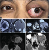
Figure 1
Figure 1
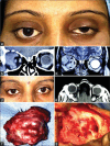
(a) Clinical photographs showing left eye proptosis and inferonasal dystopia. (b) MRI imaging, coronal cut showing a left lacrimal gland mass (adenoid cystic carcinoma) which is hypo intense on T1 sequence and extending to temporal fossa. (c) MRI imaging, axial cut showing a lacrimal gland mass (adenoid cystic carcinoma) having intermediate intensity with hyper intense foci on T2 sequence. (d and e) MRI imaging showing mild diffusion restriction within the mass on apparent diffusion coefficient (ADC) map and diffusion weighted imaging (DWI)
Figure 2
Figure 2
(a) Clinical photograph showing left eye proptosis in a case of adenoid cystic carcinoma of lacrimal gland. (b) MRI imaging, coronal cut showing a left lacrimal gland mass (adenoid cystic carcinoma) which is hypo intense on T1 sequence. The mass is compressing the adjoining lateral rectus and levatorpalpebri-superior rectus complex. (c) MRI imaging, axial cut showing a left lacrimal gland mass (adenoid cystic carcinoma) having heterogeneous hyper intense signal with hypo intense foci within it on T2 sequence. (d and e) MRI imaging showing no significant diffusion restriction within the mass on apparent diffusion coefficient (ADC) map and diffusion-weighted imaging (DWI)
Figure 3
Figure 3
(a) CT scan coronal cut showing a left lacrimal gland mass (adenoid cystic carcinoma) with specs of calcification with in it. (b-f) Histopathology images showing cribriform, Swiss cheese, basaloid with comedo necrosis, solid, and myxoid variants of adenoid cystic carcinoma, respectively. (g and h) Histopathology images showing solid variant of adenoid cystic carcinoma and perineural invasion (yellow arrow), respectively
Figure 4
Figure 4
(a and b) Pre- and post-neo-adjuvant chemotherapy clinical picture of a patient with left adenoid cystic carcinoma of lacrimal gland. Note the significant decrease in the amount of proptosis. (c and d) Pre- and post-neo-adjuvant chemotherapy radiological picture of a patient with left adenoid cystic carcinoma of lacrimal gland. Note the significant decrease in the size of mass
Figure 5
Figure 5
(a) Clincial photograph of a patient with right adenoid cystic carcinoma of the lacrimal gland. (b) CT scan coronal cuts showing an iso to hyperdense mass arising from right lacrimal gland with surrounding bony erosion. (c) MRI coronal cuts, T2-weighted sequence showing a heterogeneous intensity lesion with foci of hyper intensity arising from right lacrimal gland. (d) Clinical photograph of the same patient 1 year after excision biopsy with recurrence. (e) MRI axial cuts showing a contrast enhancing mass in the supero temporal orbit extending up to orbital excess (recurrent adenoid cystic carcinoma). (f) Gross photograph of exenterated specimen of the patient. (g) Intraoperative appearance of the exenterated socket showing multiple areas of bony erosions
Figure 6
Figure 6
Kaplan–Meier survival analysis curve (nonbasaloid vs basaloid)
Figure 7
Figure 7
Kaplan–Meier survival analysis curve (>T3 stage of TNM vs < T3 stage of TNM)
Figure 8
Figure 8
Kaplan–Meier survival analysis curve (Excision + RTVs. Exenteration ± RT)
Figure 9
Figure 9
Kaplan–Meier survival analysis curve (Excision + RTVs. Excision + RT + CT)

References

    1. Benali K, Benmessaoud H, Aarab J, Nourreddine A, El Kacemi H, El Majjaoui S, et al. Lacrimal gland adenoid cystic carcinoma: Report of an unusual case with literature review. Radiat Oncol J. 2021;39:152–8. - PMC - PubMed
    1. Ahmad SM, Esmaeli B, Williams M, Nguyen J, Fay A, Woog J, et al. American Joint Committee on Cancer classification predicts outcome of cases with lacrimal gland adenoid cystic carcinoma. Ophthalmology. 2009;116:1210–5. - PubMed
    1. Font RL, Smith SL, Bryan RG. Malignant epithelial tumors of the lacrimal gland: A clinicopathologic study of 21 cases. Arch Ophthalmol. 1998;116:613–6. - PubMed
    1. Ahmed A, Ganesan SK, Haleem S, Nicoll J. A rare case of late solitary vertebral metastasis from an adenoid cystic carcinoma of the lacrimal gland. BMJ Case Rep. 2017;2017:bcr2016218371. - PMC - PubMed
    1. Andreoli MT, Aakalu V, Setabutr P. Epidemiological trends in malignant lacrimal gland tumors. Otolaryngol Head Neck Surg. 2015;152:279–83. - PMC - PubMed

MeSH terms

LinkOut - more resources