. 2025 Dec;14(1):2562210.
doi: 10.1080/2162402X.2025.2562210. Epub 2025 Sep 25.

IL-21-reprogrammed Vδ1 T cells exert killing against solid tumors which is enhanced by CAR arming for off-the-shelf immunotherapy

Affiliations

IL-21-reprogrammed Vδ1 T cells exert killing against solid tumors which is enhanced by CAR arming for off-the-shelf immunotherapy

Ana L Portillo et al. Oncoimmunology. 2025 Dec.

Abstract

Cancer cell therapies have primarily focused on engineering autologous αβ T cells with chimeric antigen receptors (CARs), achieving clinical success against hematologic malignancies. However, their effectiveness against solid tumors is limited by challenges such as antigen escape, suppression by the metabolically hostile tumor microenvironment (TME), and manufacturing difficulties. γδ T cells are unconventional T cells with innate tumor-targeting capabilities independent of MHC class I, making them an emerging candidate for allogeneic cell therapy. While the Vδ1 T cell subset has shown promising anti-tumor killing their clinical application has been hindered by difficulties in achieving robust expansion for therapeutic use. Here, we evaluated the potential of K562 feeder cells expressing membrane-bound IL-21 (K562-mb-IL-21) to expand and activate γδ T cells from peripheral blood. Our findings show that this method preferentially expands Vδ1 T cells, resulting in an activated phenotype characterized by enhanced expression of NK cell activation receptors, innate cytotoxicity against breast and ovarian cancer cells, and sustained metabolic function in patient-derived ascites TME. When engineered with a CAR, Vδ1 T cells exhibited further enhanced anti-tumor efficacy in an immunodeficient NRG xenograft model of human ovarian cancer. These findings highlight K562-mb-IL-21 expanded peripheral blood Vδ1 T cells as a promising 'off-the-shelf' allogeneic therapy for solid tumors.

Keywords: CAR-T cells; Cell therapy; IL-21; metabolic fitness; solid tumors; γδ T cells.


Conflict of interest statement

MNK is a co-inventor of gamma delta T cell gene editing technology, he is also the co-founder and CSO of CARTx Therapeutics and owns stock in the company. All other authors declare no competing interests.

Figures

Figure 1.
Figure 1.
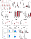
K562 mb-IL-21 feeder cells lead to Vδ1 γδ T cell expansion from PBMCs long-term. (A) Bulk PBMCs or CD3+ T cells were isolated from healthy donor peripheral blood and expanded using K562-mb-IL-21 feeder cells and IL-2 (100 U/mL) weekly. The expansion of NK cells, αβ T cells, and γδ T cell from each co-culture was tracked weekly. (B) Representative flow plots showing the percent αβ TCR+ and γδ TCR+ events of CD3+ T cells pre-expansion and at six weeks post-expansion. (C) Fold expansion of NK cells (CD3-CD56+), αβ T cells and γδ T cells in bulk PBMC co-cultures. (D) Fold expansion of αβ T cells and γδ T cells in CD3+ isolated co-cultures. (E) The proportion of NK cells, αβ T cells, and γδ T cells out of total live cells in co-cultures expanded from bulk PBMCs (F,G) the proportion of αβ TCR+ and γδ TCR+ cells out of CD3+ T cells in co-cultures expanded from bulk PBMCs (F) or from CD3+ T cells (G). (H) The percent αβ TCR+ and γδ TCR+ cells of CD3+ T cells five weeks post-expansion in bulk PBMC or CD3+ isolated T cell co-cultures. I) Representative flow plots showing the percent Vδ1, Vδ2, and Vδ1- Vδ2- γδ T cells expanded from bulk PBMCs pre-expansion and eight weeks post-expansion. The proportion of Vδ1, Vδ2, and Vδ1-Vδ2- γδ T cells in co-cultures over eight weeks of expansion was graphed. J) The percentage of Vδ1, Vδ2, and Vδ1-Vδ2- γδ T cells pre-expansion and eight weeks post-expansion. Data represent mean ± SEM of four to six biological replicates per condition. **p < 0.01, ***p < 0.001, ns, not significant (H; two-way ANOVA with Sidak’s post hoc tests, J; two-way ANOVA with Tukey’s post hoc tests).
Figure 2.
Figure 2.
Expanded γδ T cells have enhanced expression of activation markers and exhibit innate cytotoxicity against various solid tumor cell lines. (A) Representative flow plots showing expression of activation and inhibitory markers in freshly isolated or expanded γδ T cells. (B) The percent expression of activation markers and inhibitory markers was quantified by flow cytometry (n = 6). (C) Cell-mediated cytotoxicity of freshly isolated and expanded γδ T cells against OVCAR8 cells at different effector: target (E:T) ratios. The percent-specific lysis was graphed. D) The percent expression and MFI of perforin, granzyme A, and granzyme B was measured intracellularly in freshly isolated and expanded γδ T cells. (E) Cell-mediated cytotoxicity of expanded γδ T cells against OVCAR8, SKBR3, and MDA-MB-231 tumor cell lines. Graphs show the percent-specific lysis. (F-H) Expanded NK cells or γδ T cells were co-cultured with OVCAR8 or MDA-MB-231 cells at a 1:1 E:T ratio for 5 h. F) Representative flow plots of IFN-γ and CD107a expression by expanded γδ T cells and NK cells co-cultured with OVCAR8 cells. G,H) The percent expression of IFN-γ and CD107a in the effector cells after incubation with OVCAR8 (G) or MDA-MB-231(H) cells was quantified by flow cytometry. Data represent mean ± SEM of three to nine biological replicates per condition. *p < 0.05, **p < 0.01, ***p < 0.001, ****p < 0.0001 ns, not significant (B,D; paired two-tailed t-test, C; two-way ANOVA with Sidak’s post hoc tests, G; unpaired two-tailed t-test).
Figure 3.
Figure 3.
Expanded Vδ1 T cells reduce tumor burden in an OVCAR8 human ovarian cancer xenograft model. A) Expanded γδ T cells were adoptively transferred intraperitoneally (i.p.) to NRG mice two days post-injection with OVCAR8-luciferase cells. IL-2 (20,000 U/mouse) was administered every two to three days to support γδ T cell survival in vivo. Schematic shows experimental procedure. B) The tumor burden was quantified via bioluminescence at different time points. C) Tumor burden at day 21 post tumor engraftment. Radiance units are photons/sec/cm2/sr. Data represent mean ± SEM of four to six mice per group. **p < 0.01, ****p < 0.0001 (B; two-way ANOVA with Sidak’s post hoc tests, C; unpaired two-tailed t test).
Figure 4.
Figure 4.
Expanded γδ T cell cytotoxicity is not abrogated by blocking NKG2D, DNAM-1, or the γδ T cell receptor. (A) Schematic of transwell killing assay: OVCAR8 cells were seeded in the basolateral side and expanded γδ T cells were added to the apical side of a 0.4 μm transwell. After five hours, the percent live tumor cells seeded in the transwell or after direct contact with expanded γδ T cells was calculated. (B) Schematic: expanded γδ T cells were incubated with blocking antibodies for one hour followed by co-culture with tumor target cells for an additional five hours at a 4:1 effector-to-target ratio. (C, D) Relative change in specific lysis against OVCAR8 cells in the presence of blocking antibodies against NKG2D and the γδ TCR either individually (C) or in combination (D) was calculated compared to isotype controls. E) Relative change in specific lysis against MDA-MB-231 cells in the presence of blocking antibodies against NKG2D and the γδ TCR was calculated compared to isotype controls. F,G) Relative change in specific lysis against OVCAR8 (F) or MDA-MB-231 (G) cells in the presence of blocking antibodies against DNAM-1, NKG2D, and the γδ TCR was calculated compared to isotype controls. Data represent mean ± SEM of three to four biological replicates per condition. **p < 0.01, ns, not significant (A,D; unpaired two-tailed t-test, C,E-G; one-way ANOVA with Dunnett’s post hoc tests).
Figure 5.
Figure 5.
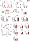
Expanded γδ T cells sustain their cytotoxicity and metabolic function in the malignant ascites tumor microenvironment. A) Representative flow plots showing expression of CD71, CD98 and Glut1 in fresh and expanded γδ T cells. The percent and MFI expression of CD71, CD98 and Glut1 was measured by flow cytometry. B) Schematic showing incubation of expanded γδ T cells in media or the ascites tumor microenvironment (AscTME) for three days prior to cytotoxicity and metabolism assessment. C) Expanded γδ T cell-mediated cytotoxicity against OVCAR8 cells was measured at a 1.25 E:T ratio after incubation in media or AscTME. D) Cell surface expression of CD71, CD98 and Glut1 on expanded and freshly isolated γδ T cells was measured by flow cytometry after incubation in media or the AscTME. Relative change in expression compared to media only controls was quantified. E) Representative measures of ECAR in one donor. F) Quantified basal glycolysis, glycolytic capacity, and glycolytic reserve. G) Representative measures of OCR in one donor. H) Quantified basal oxidative phosphorylation (OxPhos), maximal OxPhos, spare respiratory capacity (SRC), and mitochondrial linked-ATP production. Data represent mean ± SEM of four to seven biological replicates per condition. *p < 0.05, ***p < 0.001, ****p < 0.0001. ns, not significant (A,C,F,H; paired two-tailed t-test, D; two-way ANOVA with Sidak’s post hoc tests).
Figure 6.
Figure 6.
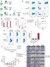
Expanded Vδ1 T cells expressing a HER2-specific CAR show enhanced anti-tumor functions against HER2-overexpressing tumors. A) Schematic depicting the generation of HER2 CAR/AAVS1 KO Vδ1 T cells and wild type Vδ1 T cell controls using CRISPR/Cas9 electroporation and AAV transduction. B) Flow cytometry plots showing expression of the HER2 CAR on expanded γδ T cells. C) The percent expression of the HER2 CAR was measured every seven days by flow cytometry over 21-days of expansion. D) Cell-mediated cytotoxicity of HER2 CAR/AAVS1 KO-, WT-Electroporated-, or WT-AAVS1 KO- Vδ1 T cells against HER2+ SKBR3 or HER2- OVCAR8 tumor cells at the indicated effector-to-target (E:T) ratios. The percent specific lysis was graphed. E) The cell surface expression of CD107a (degranulation) on HER2 CAR/AAVS1 KO-, WT-Electroporated-, or WT-AAVS1 KO- Vδ1 T cells were measured at baseline and after co-culture with HER2+ SKBR3 tumor cells after 5 hours at a 1:1 E:T ratio. The relative expression of CD107a compared to baseline was calculated and graphed. Data represent mean of technical replicates from one biological donor. F) NRG mice were implanted intraperitoneally (i.P.) with 0.50x10⁶ SKOV3-luciferase cells followed by 10x106 HER2 CAR/AAVS1 KO-, WT-Electroporated-, or WT-AAVS1 KO- Vδ1 T cells two days after tumor engraftment. IL-2 (20,000 U/mouse) was given every two to three days to support γδ T cell survival. The tumor burden was quantified via bioluminescence at different time points and graphed. Data represent mean ± SEM of three to seven mice per group. **p < 0.01, ****p < 0.0001. ns, not significant (F; two-way ANOVA with Tukey’s post hoc tests).

References

    1. Liu Y, Zhang C.. The role of human γδ T cells in anti-tumor immunity and their potential for cancer immunotherapy. Cells. 2020;9(5):1206. doi: 10.3390/CELLS9051206. - DOI - PMC - PubMed
    1. Gentles AJ, Newman AM, Liu CL, Bratman SV, Feng W, Kim D, Nair VS, Xu Y, Khuong A, Hoang CD, et al. The prognostic landscape of genes and infiltrating immune cells across human cancers. Nat Med. 2015;21(8):938–22. doi: 10.1038/nm.3909. - DOI - PMC - PubMed
    1. Hinz T, Wesch D, Halary F, Marx S, Choudhary A, Arden B, Janssen O, Bonneville M, Kabelitz D. Identification of the complete expressed human TCR V gamma repertoire by flow cytometry. Int Immunol. 1997;9(8):1065–1072. doi: 10.1093/intimm/9.8.1065. - DOI - PubMed
    1. Deusch K, Lüling F, Reich K, Classen M, Wagner H, Pfeffer K. A major fraction of human intraepithelial lymphocytes simultaneously expresses the γ/δ T cell receptor, the CD8 accessory molecule and preferentially uses the Vδ1 gene segment. Eur J Immunol. 1991;21(4):1053–1059. doi: 10.1002/eji.1830210429. - DOI - PubMed
    1. Fonseca S, Pereira V, Lau C, Teixeira MDA, Bini-Antunes M, Lima M. Human peripheral blood gamma delta T cells: report on a series of healthy Caucasian Portuguese adults and comprehensive review of the literature. Cells. 2020;9(3):729. doi: 10.3390/cells9030729. - DOI - PMC - PubMed

MeSH terms