. 2025 Sep 25;17(1):102.
doi: 10.1186/s13073-025-01538-1.

EMB is essential for enteric nervous system development mediated by PI3K signaling

Affiliations

EMB is essential for enteric nervous system development mediated by PI3K signaling

Zhi Li et al. Genome Med. .

Abstract

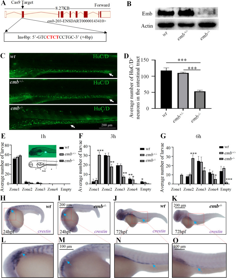
Background: The enteric nervous system (ENS), which arises from enteric neural crest cells (ENCCs), plays important roles in many aspects of gastrointestinal tract function, including motility, secretions, blood flow and hormone release. Defects in ENS development could lead to a broad range of disorders, including Hirschsprung's disease (HSCR), which is characterized by missing nerve cells in the distal segment of the colon. Here, we identify EMB as an evolutionarily conserved regulator of ENS development.

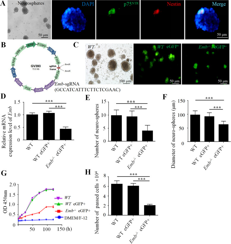
Methods: We first examined EMB expression in human and mouse intestines using scRNA-seq data and immunofluorescence staining. To investigate its role in ENS development, we constructed Emb-knockout zebrafish and mouse models. To explore the underlying mechanisms, we focused on ENCCs and analyzed their proliferation and migration using migration assays in explant guts and organoid cultures. Finally, we assessed rare EMB variants in a cohort of HSCR patients.

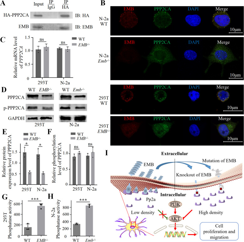
Results: In zebrafish, loss of emb leads to a decrease number of enteric neurons and impaired intestinal transit ability. In mice, knockout of Emb causes HSCR-like phenotypes and defects. In vitro experiments, including explant mouse gut and organoid cultures, show that EMB is required for both the proliferation and migration of ENCCs. Mechanistically, EMB binds to and recruits the phosphatase complex PP2A to the cellular membrane to facilitate the activation of PI3K-AKT pathway, thereby promoting ENCCs development. Indeed, application of PI3K or AKT agonists partially restores the ENS developmental defects in zebrafish emb mutants. Furthermore, rare variants of EMB may potentially contribute to the pathology of HSCR in humans.

Conclusions: EMB is required for ENS development by regulating the proliferation and migration of the ENCCs. Mechanistically, EMB recruits PP2A to the cell membrane, reducing cytoplasmic dephosphorylation activity and promoting the activation of the PI3K signaling pathway.

Keywords: EMB; Enteric nervous system; Hirschsprung’s Disease; Neural crest cell; PI3K pathway.


Conflict of interest statement

Declarations. Ethics approval and consent to participate: All protocols involving human sample collection in this study were approved by the Ethics Committee of Tongji Medical College of Huazhong University of Science and Technology (Ethical approval number: 2020-S226, 2021-S033). All relevant parts of the research were conducted in accordance with the Declaration of Helsinki, and written informed consent was obtained from all participants or their legal guardians. Experiments about zebrafish or mice were approved by the Scientific Ethics Committee of Huazhong Agricultural University (Ethical approval number: HZAUMO-2017-048). Consent for publication: Not applicable. Competing interests: The authors declare no competing interests.

Figures

Fig. 1
Fig. 1
Loss of emb causes ENS defects in zebrafish. A, Generation of targeted emb mutant in Zebrafish. A 4 bp insertion in Exon 2 was introduced through the CRISPR/Cas9 system. B, Expression of the Emb protein is disrupted in different genotypes determined by western blots. C, The enteric neurons at 5 dpf zebrafish larva of wild type (wt), heterozygous and homozygous emb mutants are labeled by HuC/D and white arrows point to the most posterior margin where the enteric neurons migrated to. D, Quantification of the numbers of HuC/D positive cells in the intestinal tract, ***, p < 0.001, one-way ANOVA with Bonferroni post-hoc, N = 10 for each group. E-G, Quantification of the number of larvae in each group based on the anterior extent of tracer. “empty” means fluorescent tracers are completely evacuated in the intestine. Statistical significances compared to wt are labeled on top of each bar, *, p < 0.05; **, p < 0.01; ***, p < 0.001, one-way ANOVA with Bonferroni post-hoc, N = 60. H-K, Lateral view of in situ hybridization results of 24 hpf and 72 hpf larvae using crestin probe, blue arrows point to migrating neural crest cells. L-O, Locally enlarged images of the H–K images (Red dashed box)
Fig. 2
Fig. 2
Loss of Emb causes HSCR-like phenotypes in mice. A, Using CRISPR/Cas9 system, a 13-bp frameshift deletion in exon 3 of Emb was induced. B, Emb mRNA expression is diminished in Emb-/- mice determined by quantitative real-time PCR. C, The EMB protein expression levels in different genotypes were determined by Western blotting, and the expression levels were quantified in (D). E, The percentage of each genotype in three different crossing scenarios. ***, p < 0.001, one-way ANOVA with Bonferroni post-hoc, N = 3. F, Abdominal anatomy of wild type, Emb+/- and Emb-/- mice. G, Isolated colorectal from the distal rectum to the ileocecal part, note the dry stool filled and dilated colon of the Emb-/- mice. H, Immunofluorescence staining of 6-week mouse colons; the entire colon is rolled into a donut-like shape along the longitudinal axis, and then embedded and processed into paraffin sections; the right panels are zoom-ins of the distal colon. Green, Tuj1; blue, DAPI. The red arrows indicate the enteric neurons farthest from the proximal colon, and the distance from the"red arrow"to the distal colon represents the aganglionic segment; PC, proximal colon; DC, distal colon; Quantification results are presented in Additional file 2: Fig.S5 G. I, Immunofluorescence staining of the cross sections of 2-day neonatal mouse colons. Red, Brdu; green, Tuj1; blue, DAPI. Quantification results are presented in Additional file 2: Fig.S5 H
Fig. 3
Fig. 3
EMB is required for ENCC proliferation and migration in vitro. A, Isolated and cultured ENCCs identified by immunofluorescence double labeling of p75 (green) and Nestin (red), the nuclei are labeled with DAPI (blue). B, Schematic overview of the lentiviral construct used for Emb gene knockout in ENCCs. C, Lentivirus infection is confirmed by GFP fluorescence at day 6. D, Relative mRNA expression level of Emb in neurospheres. E, Numbers of the neurospheres are reduced upon knockout of Emb. F, Average diameter of the neurospheres is reduced upon knockout of Emb. G, Cell growth curve in the proliferation assay showing that loss of Emb affects ENCC proliferation. H, The migration ability, accessed by numbers of cells passed the matrix in the transwell assay, is affected by loss of Emb. **, p < 0.01, ***, p < 0.001, one-way ANOVA with Bonferroni post-hoc, N = 6
Fig. 4
Fig. 4
Emb is required for the migration of the ENCCs in gut explant. A, Schematic illustrating the ENCC migration assay in mouse gut explants. B Using the transgenic mice with ENCCs labeled by genetically encoded tdTomato, and the ENCCs were marked by Sox10 (red) in E12.5d mice. C, Relative density of ENCCs behind the leading ENCCs, NS, non-significance, one-way ANOVA with Bonferroni post-hoc, N = 4. D, Representative images of the in vitro ENCC migration assay, ENCCs are labeled with P75NTR (red) and Sox10 (green). E, Quantification methods of the in vitro migration assay, the membranes were divided into five regions based on the distance from the margin of the explant and the numbers of Sox10 positive cells in each region were counted and quantified. F and G, Mean migration distance of Sox10+ cells and migration distance of the furthest migrating Sox10+ cell in different groups; **, p < 0.01; ***, p < 0.001, one-way ANOVA with Bonferroni post-hoc, N = 3. H and I, Polar histograms representing the trajectories of the cells which were 150 μm from the most caudal cell at 40-min intervals in WT (H) and Emb-/- explants of E12.5d hindgut (I). The trajectories are determined by drawing a straight line from the position of cell to its position 40 min previously, with the 0 degree as the rostrocaudal axis of the gut (27 cells were examined). J, Quantification of the average net speed by measuring the distance between the location of the wave front at the beginning of the time lapse sequence and its location at the end of the sequence. ***, p < 0.001, two-tailed Student’s T test, N = 3
Fig. 5
Fig. 5
Emb regulates ENS development through the activation of PI3K pathway in zebrafish. A-C, Validation of the efficiency of the PI3K/AKT agonists (740-Y-P and SC79) in wild type zebrafish. D, Western blots of total and phosphorylated PI3K and AKT of 5 dpf zebrafish larvae, before and after exposure of agonists, the protein levels are quantified in (E and F). Representative images of immunofluorescence staining to assess the number of enteric neurons (G, HuC/D) and the levels of cell proliferation (H, p-H3). In Figure G, the dashed box in the left panel marks the most distal enteric neurons positive for HuC/D; the right panel shows a magnified view of the boxed region. In Figure H, the dashed box in the left panel indicates a representative region with high PH3 expression; the right panel shows a magnified view of the boxed region. The quantification results are presented in I and J. *, p < 0.05; **, p < 0.01; ***, p < 0.001, one-way ANOVA with Bonferroni post-hoc, N = 10
Fig. 6
Fig. 6
EMB activates PI3K pathway by recruiting PP2A to the cell membrane. A, Co-IP between EMB and PPP2CA in N-2a cells. B, Immunofluorescence staining of EMB (red) and PPP2CA (green) in WT or Emb-/- N-2a or 293 T cells. The yellow puncta indicate co-localization (C), Ppp2ca mRNA expression levels determined by RT-qPCR. D, Upon fractionation, western blots of total and phosphorylated membrane-associated Ppp2ca in 293 T or N-2a cells. E and F, Quantification of the protein levels in panel D. G and H, The Serine/threonine phosphatase activities of PP2A enzymes in the cytoplasmic fraction. ns, not significance; *, p < 0.05; ***, p < 0.001. I, A working model of EMB function in PI3K signaling

References

    1. Heuckeroth RO. Hirschsprung disease - integrating basic science and clinical medicine to improve outcomes. Nat Rev Gastroenterol Hepatol. 2018;15(3):152–67. - PubMed
    1. Furness JB. The enteric nervous system and neurogastroenterology. Nat Rev Gastroenterol Hepatol. 2012;9(5):286–94. - PubMed
    1. Huang T, Hou Y, Wang X, Wang L, Yi C, Wang C, et al. Direct Interaction of Sox10 With Cadherin-19 Mediates Early Sacral Neural Crest Cell Migration: Implications for Enteric Nervous System Development Defects. Gastroenterology. 2022;162(1):179-92.e11. - PubMed
    1. Zhou B, Feng C, Sun S, Chen X, Zhuansun D, Wang D, et al. Identification of signaling pathways that specify a subset of migrating enteric neural crest cells at the wavefront in mouse embryos. Dev Cell. 2024;59(13):1689-706.e8. - PubMed
    1. Landman KA, Simpson MJ, Newgreen DF. Mathematical and experimental insights into the development of the enteric nervous system and Hirschsprung’s disease. Dev Growth Differ. 2007;49(4):277–86. - PubMed

Substances

LinkOut - more resources