

Giant Uterine Leiomyoma Causing Severe Hemodynamic Instability
- PMID: 41000558
- PMCID: PMC12456486
- DOI: 10.1097/og9.0000000000000059
Giant Uterine Leiomyoma Causing Severe Hemodynamic Instability
Abstract
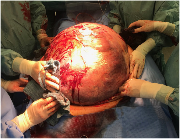
Background: Uterine leiomyomas occasionally manifest as giant leiomyomas, displaying exceptional sizes within the uterus and abdominal cavity. These massive growths, although rare because of increased patient awareness, can severely distort pelvic structures and pose life-threatening complications by compressing adjacent organs. We report a case of a patient who presented with severe hemodynamic instability, cardiac arrest, and multiorgan failure caused by a giant 54.8-kg uterine leiomyoma, representing the heaviest weight ever reported in the literature in surviving patients so far.
Case: This article documents a case of a 56-year-old woman with a giant uterine leiomyoma in critical condition characterized by multiorgan failure and acute kidney injury necessitating urgent surgical intervention. A laparotomy and subsequent total abdominal hysterectomy were performed. Histopathologic examination revealed a giant uterine leiomyoma. Postoperatively, the patient underwent intensive care management followed by rehabilitation, exhibiting significant clinical recovery.
Conclusion: Managing giant uterine leiomyomas demands a multidisciplinary approach, emphasizing tailored treatment strategies and specialized surgical expertise. The case report describes one of the largest leiomyomas in the literature, underscoring its rarity, and highlights the significance of timely diagnosis and appropriate interventions.
© 2025 The Authors. Published by Wolters Kluwer Health, Inc. on behalf of the American College of Obstetricians and Gynecologists.
Figures


References
Publication types
LinkOut - more resources
Full Text Sources