[Preprint]. 2025 Sep 17:2025.09.14.676119.
doi: 10.1101/2025.09.14.676119.

CNS-Tau Specific Antibodies Illuminate Disease Signatures Across Tauopathies

Affiliations

CNS-Tau Specific Antibodies Illuminate Disease Signatures Across Tauopathies

Lei Liu et al. bioRxiv. .

Abstract

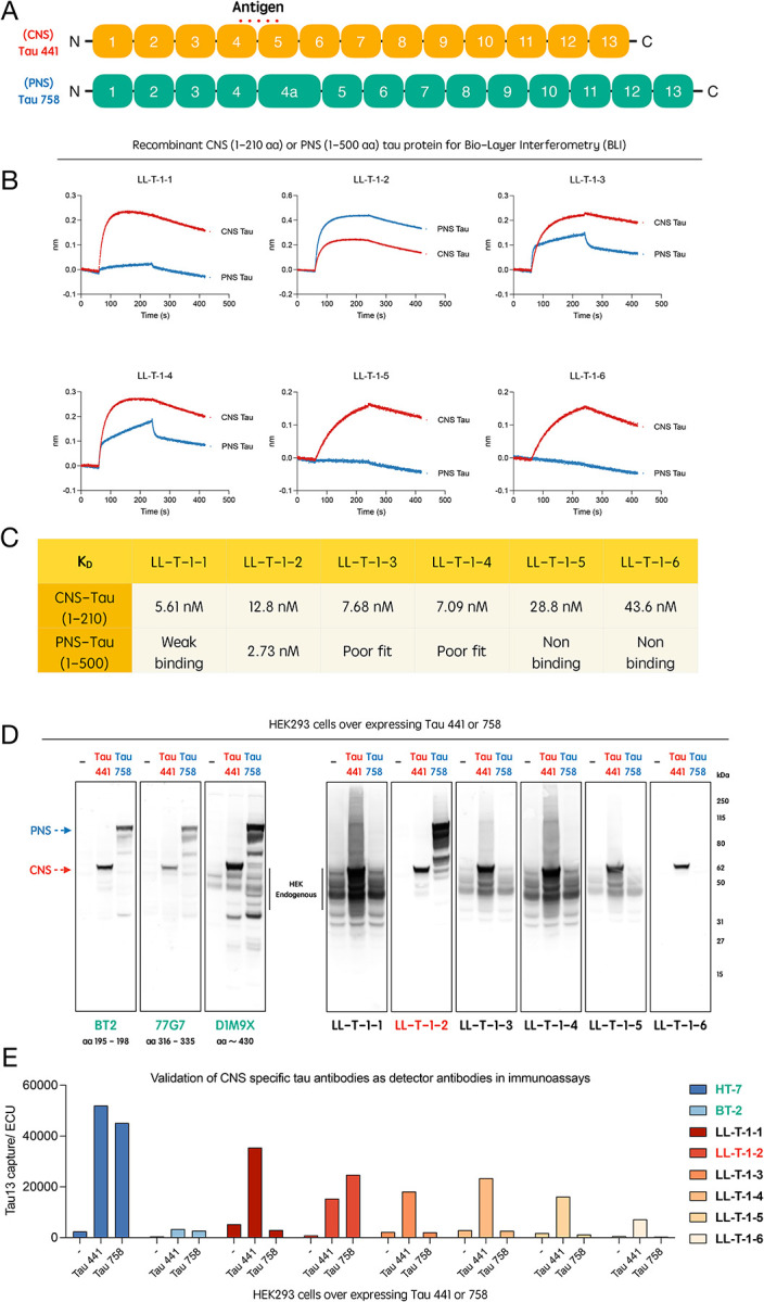
Background: Alternative splicing of the MAPT gene produces distinct tau isoforms in the central and peripheral nervous systems (CNS and PNS), yet their respective biological and pathological roles remain poorly understood. Recent studies suggest that CNS-tau may play a key role in amyloid-β associated neurodegeneration in Alzheimer's disease (AD), but the absence of isoform-specific tools has limited both mechanistic insight and biomarker development. We aimed to develop and validate CNS-tau-specific monoclonal antibodies and assess their utility in neuropathology and fluid-based biomarker assays across AD and primary tauopathies.

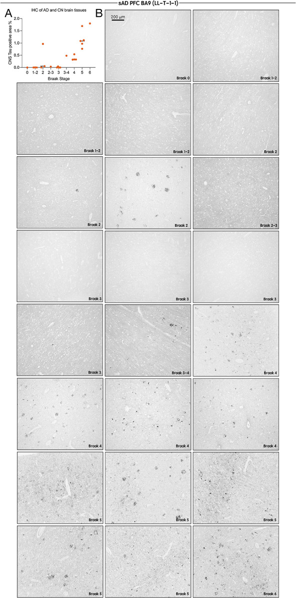
Methods: We generated six recombinant rabbit monoclonal antibodies targeting a CNS-tau-specific sequence encoded by exons 4 and 5. Specificity and affinity were evaluated via biolayer interferometry, immunoblotting, and tau-expressing HEK293 models. The lead clone, LL-T-1-1, was tested in postmortem brain sections from AD (n = 23), progressive supranuclear palsy (PSP, n = 3), and corticobasal degeneration (CBD, n = 3). A second clone, LL-T-1-5, was optimized for use in plasma assays via an ultrasensitive nanoneedle platform, LoD < 1 pg/ml.

Results: LL-T-1-1 showed nanomolar affinity for CNS-tau and no cross-reactivity with PNS-tau. It selectively labeled dystrophic neurites in AD and all hallmark tau lesions in PSP and CBD without antigen retrieval. LL-T-1-5-based plasma assays revealed CNS-tau levels significantly correlated with cognitive scores (MMSE and QDRS) and differentiated impaired from unimpaired individuals.

Conclusions: CNS-tau-specific antibodies LL-T-1-1 and LL-T-1-5 provide new tools for neuropathology and fluid biomarker development across tauopathies.

Keywords: Alzheimer’s disease; CNS-specific tau isoform; neurodegenerative disease.


Conflict of interest statement

Declaration of Interests Luke Slominski and Qimin Quan are employees of NanoMosaic, Inc. DJS is a director of Prothena Biosciences and an ad hoc consultant to Eisai and Roche. All other authors have nothing to disclose.

Figures

Figure 1.
Figure 1.. Generation and validation of CNS-tau–specific antibody series LL-T-1.
(A) Schematic representation of the tau protein, highlighting the epitope region unique to CNS-tau isoforms (junction sequence between protein encoded by exons 4 and 5). (B) Sensorgrams from biolayer interferometry (BLI) showing binding kinetics of LL-T-1 antibodies to recombinant CNS- and PNS-tau proteins. (C) Calculated equilibrium dissociation constants (Kd) calculated from BLI data for all six antibodies against CNS- and PNS-tau. (D) Immunoblots of lysates from HEK293 cells transfected with full-length CNS-tau (Tau-441) or PNS-tau (Tau-758), probed with commercial tau antibodies or LL-T-1 clones. (E) Isoform-selective immunoassays using Tau-13 as the capture antibody and commercial antibodies or LL-T-1 clones as detectors.
Figure 2.
Figure 2.. LL-T-1–1 preferentially labels dystrophic neurites in AD and robustly detects classic tau lesions in PSP and CBD brains.
(A) High-magnification images of LL-T-1–1 immunostaining in Alzheimer’s disease (AD) brain sections reveal abundant labeling of dystrophic neurites (1), neurofibrillary tangles (2), and corpora amylacea (3) (red labels). (B) High-magnification immunofluorescence images showing LL-T-1–1 (cyan) and AT8 (purple) co-staining. LL-T-1–1 and AT8 co-label dystrophic neurites, while AT8 additionally stains neurofibrillary tangles that are not consistently recognized by LL-T-1–1, as highlighted by the white circle. (C) LL-T-1–1 immunostaining in progressive supranuclear palsy (PSP) brain sections demonstrate robust labeling of globose tangles (4), tufted astrocytes (5), and oligodendroglial coiled bodies (6) (red labels). (D) In corticobasal degeneration (CBD) brain sections, LL-T-1–1 highlights ballooned neurons (7), astrocytic plaques (8), and neuropil threads (9) (red labels).
Figure 3.
Figure 3.. LL-T-1–1 labeling of tau pathology in AD brains reflects Braak stage progression.
(A) Quantification of LL-T-1–1 immunohistochemical staining densities in the BA9 region across all Alzheimer’s disease (AD) cases (n = 23). (B) Low-magnification images of LL-T-1–1 immunohistochemistry in the BA9 region of AD brain sections grouped by Braak stages 0–6. LL-T-1–1–positive tau pathology increases with advancing Braak stage, consistent with the characteristic spatial progression of tau pathology in AD.
Figure 4.
Figure 4.. Plasma CNS-tau levels correlate with cognitive impairment.
(A) Schematic of the nanoneedle array integrated into a 96-well plate format. (B) High-magnification dark-field image of the nanoneedle surface prior to assay. (C) Illustration of the sandwich immunoassay, with LL-T-1–5 antibody immobilized on each nanoneedle for target capture, followed by detection using labeled secondary antibodies. (D) Representative post-assay image showing colorimetric signal changes on the nanoneedle array compared to (B). (E) Correlation of plasma CNS-tau levels with (left to right) age, QDRS score, and MMSE score. Pearson’s correlation coefficients were used for statistical analysis. (F) Cross-sectional comparisons of plasma CNS-tau levels between cognitively impaired and unimpaired individuals, classified by MMSE (< 24) or QDRS (> 2). Group differences were assessed using the non-parametric Mann–Whitney U test. ***p < 0.001.

References

    1. Liu L, Kwak H, Lawton TL, et al. An ultra-sensitive immunoassay detects and quantifies soluble Aβ oligomers in human plasma. Alzheimer’s & Dementia. 2022;18(6):1186–1202. doi: 10.1002/alz.12457 - DOI
    1. Gonzalez-Ortiz F, Turton M, Kac PR, et al. Brain-derived tau: a novel blood-based biomarker for Alzheimer’s disease-type neurodegeneration. Brain. 2023;146(3):1152–1165. doi: 10.1093/brain/awac407 - DOI - PMC - PubMed
    1. Gonzalez-Ortiz F, Kirsebom BE, Contador J, et al. Plasma brain-derived tau is an amyloid-associated neurodegeneration biomarker in Alzheimer’s disease. Nat Commun. 2024;15(1):2908. doi: 10.1038/s41467-024-47286-5 - DOI - PMC - PubMed
    1. Salvadó G, Horie K, Barthélemy NR, et al. Disease staging of Alzheimer’s disease using a CSF-based biomarker model. Nat Aging. 2024;4(5):694–708. doi: 10.1038/s43587-024-00599-y - DOI - PMC - PubMed
    1. Bellier J, Cai Y, Alam SM, et al. Uncovering elevated tau TPP motif phosphorylation in the brain of Alzheimer’s disease patients. Alzheimer’s & Dementia. 2024;20(3):1573–1585. doi: 10.1002/alz.13557 - DOI

Publication types

LinkOut - more resources