. 2025 Sep 4;5(9):4321-4336.
doi: 10.1021/jacsau.5c00694. eCollection 2025 Sep 22.

Stabilizing a Native Fold of Alpha-Synuclein with Short Helix-Constrained Peptides

Affiliations

Stabilizing a Native Fold of Alpha-Synuclein with Short Helix-Constrained Peptides

Richard M Meade et al. JACS Au. .

Abstract

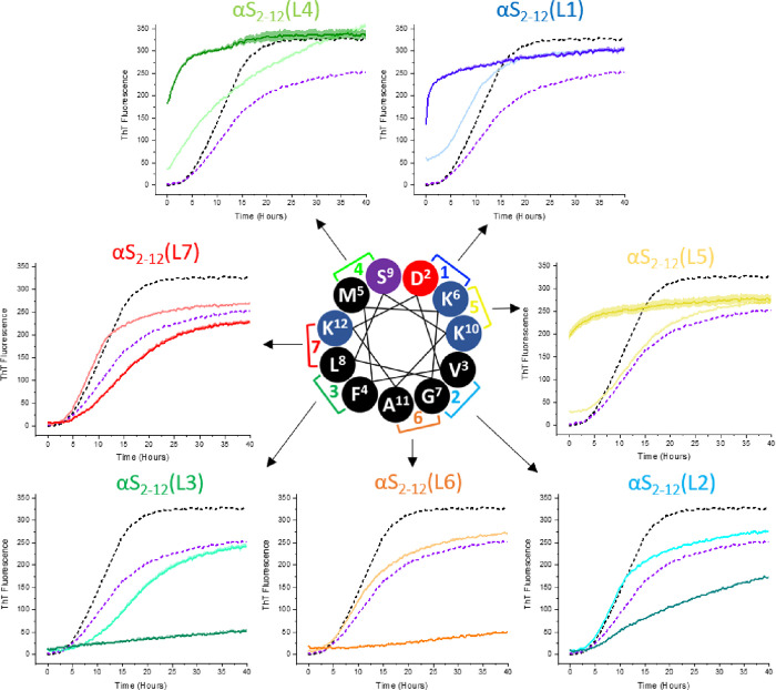
Preventing the aggregation of α-synuclein (αS) into toxic oligomers and conformers is a major therapeutic goal in conditions such as Parkinson's disease and Lewy body dementia. However, the large intracellular protein-protein interfaces within such aggregates make this a challenging target for small molecule approaches or biologics, which often lack cell permeability. Peptides occupy a suitable middle ground and are increasingly being explored as preventative treatments. We previously showed that the N-terminal lipid binding region (αS1-25) inhibits αS aggregation. Building on this, we designed a series of N- and C-terminal truncations to systematically reduce the peptide length, enabling a 56% downsizing (i.e., truncating 92% of the full-length αS protein), to identify the smallest functional unit capable of binding αS and potently blocking its aggregation and toxicity. We next introduced seven systematic i → i + 4 helix constraints to assess impact on (i) α-helicity, (ii) aggregation inhibition, (iii) serum stability, (iv) neuronal uptake, and (v) phenotypic rescue. This work maps key amphipathic features and identifies residues that are critical for αS engagement and inhibitory activity. The most effective helix-constrained peptide, αS2-12(L6), showed marked improvements across all metrics and represents a strong candidate for further therapeutic development.

Keywords: Parkinson’s disease; amyloid aggregation; lipid induced aggregation; lipid vesicles; peptide.


Figures

1
1
N-terminal fragments of αS1–140 retain lipid binding and lipid-induced aggregation inhibition properties. Adapted from Meade et al. (A) αS1–140 is an IDP in solution but adopts an α-helical conformation upon lipid binding (PDBID = 1XQ8 − ) (B) Circular dichroism confirms α-helicity of αS1–140 in the presence of DMPS SUVs. (C) A truncated N-terminal fragment of αS1–25 (82% of αS1–140 deleted) retains comparable lipid-induced helicity. (D) ThT aggregation assay shows that αS1–25 inhibits lipid induced aggregation of αS1–140 in the presence of DMPS SUVs.
2
2
Lipid binding properties of αS1–25 truncations assessed by circular dichroism (A) Table summarizing N- and C- terminal truncations of the parent αS1–25 peptide. (B) CD spectra showing lipid binding of 20 μM αS1–25 in the presence of 1000 μM DMPS vesicles (C) C-terminal truncations (removal of up to 10 residues) do not significantly disrupt lipid binding, as assessed by CD. (D) N-terminal truncations of αS1–25. (E) lipid binding of N- and C-terminal truncations of αS1–25 (F) Further truncation of αS1–15 to αS2–12 results in a peptide that retains lipid-binding capacity. (G) Peptide αS2–12 adopts a random coil structure in aqueous buffer (dotted line; 8.1% fractional helicity), which shifts to a more helical conformation (solid line; 26.2% fractional helicity) upon interaction with DMPS SUVs. All CD spectra represent the mean of three independent measurements.
3
3
Lipid binding and helicity of αS2–12 lactams analogues assessed by circular dichroism (A) Seven lactam-constrained variants of αS2–12 were synthesized, each featuring a single (i → i + 4) side-chain linkage at a different helical turn position (B) CD spectra of each peptide (20 μM; color gradient blue to red) were recorded in the absence (dotted lines) and presence (solid lines) of 1000 μM DMPS vesicles. Full-length αS1–25 (black trace) is included for reference. (C) Table summarizing fractional helicity of each peptide with and without DMPS SUVs. Among the lactams, αS2–12 (L6) exhibited the highest helicity in both the absence (31.4%) and presence (49.3%) of lipid vesicles, representing a 23% increase in helicity relative to the unconstrained αS2–12 linear peptide. All spectra represent the mean of three independent measurements.
4
4
Lactam scan identifies optimal constraint positions for inhibiting αS1–140 aggregation (A) ThT kinetic aggregation assays were performed in 20 mM phosphate buffer (pH 6.5) at 30 °C, using 100 μM αS1–140, 50 μM ThT, and 50 μM DMPS SUVs. αS2–12 lactam analogues (L1-L7) were tested as inhibitors at two concentrations: 100 μM (light traces) and 500 μM (dark traces). Aggregation of αS1–140 alone is shown as a black dashed line; linear, unconstrained αS2–12 at 500 μM is shown as a purple dashed line. Lactams αS2–12(L6) and αS2–12(L3) demonstrated the greatest inhibition of αS1–140 aggregation. In contrast, lactams bridging the hydrophilic face of the helix (L1, L4, L7)) appear to enhance aggregation. Peptides constrained on the hydrophobic face (L2, L3 and L6) were the most effective at suppressing DMPS-induced aggregation, highlighting the importance of helix face orientation in modulating activity. Data shown as mean ± standard error (n = 3).
5
5
αS2–12(L6) is a potent, dose-dependent inhibitor of αS1–140 aggregation (A) ThT kinetic assays were performed with 100 μM αS1–140, 50 μM ThT and 50 μM DMPS in 20 mM phosphate buffer (pH 6.5) at 30 °C, in the presence of increasing concentrations (0–100 μM) of αS2–12(L6). Aggregation is progressively suppressed with increasing inhibitor concentration, with complete inhibition observed at 100 μM. Data shown as mean ± standard error (n = 3). (B) TEM images from the same end point ThT assays reveal fibril structures in the absence of αS2–12(L6) (red) and substantial suppression of fibril formation in its presence (orange). (C) SDS-PAGE of protein samples collected at the ThT end point, following PICUP, shows a reduction in oligomeric αS1–140 species with increasing concentrations of αS2–12(L6). (D) Densitometric analysis of SDS-PAGE band intensities using ImageJ confirms a dose-dependent decrease in higher-order oligomer bands (bands c–f), correlating with increasing αS2–12(L6) concentration.
6
6
αS2–12(L6) has no effect on the agitation induced aggregation of αS1–140. (A) ThT kinetics of 100 μM αS1–140 and a single 3 mm glass bead to agitate the solution presented no change in aggregation with increasing concentrations of αS2–12(L6) (0–1000 μM). (B) Aggregates formed presented straight fibrils, consistent with those previously reported with αS1–140 aggregation. Data shown as mean ± standard error (n = 3).
7
7
Solution NMR structure of αS2–12(L6). (A) Lowest-energy structure of αS2–12(L6). (PDB 8OL8), showing an amphipathic α helix with side chains colored by chemical character: positively charged (blue), negatively charged (red), polar (purple), and the lactam constraint (orange). (B) Ribbon ensemble of the 20 lowest energy conformers generated from the final structure calculation, demonstrating high structural convergence. (C) Solvent-accessible surface representation of αS2–12(L6), viewed along the helical axis from the N-terminus, illustrates the distribution of exposed side chains and the amphipathic nature of the helix. The helical form comprises approximately three turns, with each position in the helical wheel occupied. The presence of an N-terminal Asp (D) and a C-terminal Lys (K), may further stabilize the peptide.
8
8
1H–15N HSQC spectra of αS1–140 in the presence of αS2–12(L6) over time. HSQC spectra were acquired for 100 μM 15N-labeled αS1–140 with 50 μM DMPS in 20 mM sodium phosphate buffer (pH 6.5) at 30 °C (A) Time (t) = day 0 spectrum of αS1–140 alone, showing well-resolved cross-peaks consistent with a disordered monomer. (B) Spectrum of αS1–140 at day 0 in the presence of αS2–12L6 at a 1:1 molar ratio, showing spectral features comparable to those of panel A. (C) Spectrum of αS1–140 alone after 6 days of incubation. Aggregated material was removed by centrifugation prior to acquisition; the remaining soluble protein displays substantial peak loss and broadening, consistent with aggregation. (D) Spectrum of αS1–140 with αS2–12(L6) after 6 days of incubation. The preservation of cross-peaks similar to those observed at day 0 indicates that αS2–12(L6) protects monomeric αS1–140 from aggregation or degradation under these conditions.
9
9
Lactamization of αS2–12(L6) enhances peptide stability in human serum. Serum stability was assessed by incubating αS2–12 (linear) and αS2–12(L6) (lactamized) in human serum at 37 °C. Peptide concentration was quantified by analytical HPLC at selected time points and normalized to t = 0. After 5 h, 96% of the linear αS2–12 was degraded, whereas only 14% of the lactamized αS2–12(L6) had been lost, indicating substantially increased protease resistance conferred by the lactam constraint. Data represent the mean of three independent experiments; error bars indicate the standard error of the mean.
10
10
Peptide uptake and cytocompatibility in SH-SY5Y cells. (A) Representative fluorescence microscopy images of SH-SY5Y cells treated with peptide L6 (10 and 20 μM) for 33 h, showing intracellular peptide uptake. Untreated cells are shown for comparison. Nuclei are stained with NucBlue (blue), cytoplasm with CellMask (red), and peptides are visualized in yellow (scale bar = 50 μm). Images shown are z-projections spanning 8 μm, capturing all in-focus fluorescence signals across the stack. (B) Quantification of intracellular uptake, represented as the number of RhoB-labeled peptide puncta relative to local background signal at each location, indicating peptide internalization. (C) Cytotoxicity of peptides αS2–12, αS2–12(L6) (constrained), and αS2–12L6 (linear) assessed by Alamar Blue and ToxiLight assays in SH-SY5Y cells. Viability is expressed relative to that of untreated controls and Triton X-100 (Tx-100) as a positive control. No significant toxicity was observed across the 0–20 μM concentration range. Each data point represents the mean of at least three technical replicates, each from independent experimental repeats involving separate platings of the SH-SY5Y cells. Note: The SH-SY5Y cells used in this study expressed low endogenous levels of αS.
11
11
αS2–12­(L6) treatment rescues disease phenotype in C. elegans expressing α-synuclein–YFP. (A) NL5901 worms expressing α-synuclein–YFP (αS–YFP) in body wall muscle were treated with αS2–12(L6) at the L4 larval stage. Mobility was assessed by thrashing frequency on Day 5 (D5) of adulthood. Worms were redosed on adult day 7 (D7), and inclusion formation was assessed via fluorescence microscopy on adult day 13 (D13). (B) On D13, NL5901 worms displayed prominent αS–YFP fluorescent inclusions along the body wall in the absence of peptide, while control N2 worms showed no fluorescence (scale bar = 200 μm, 5 worms analyzed per condition). (C) Thrashing assays performed on D5 revealed that peptide treatment had no adverse effect on N2 wild-type worms. In contrast, NL5901 worms showed significantly reduced motility (P < 0.01) without treatment, which was rescued in a dose-dependent manner by αS2–12(L6). (D) Peptide treatment significantly reduced αS–YFP inclusion burden in NL5901 worms on D13 (P < 0.01) (5 worms analyzed per condition). (E) Representative images of worm heads showing αS–YFP fluorescence with and without peptide treatment. A reduction in the level of fluorescent inclusions is observed in peptide-treated worms. Full-body images are provided in Supplementary Figure 6.

References

    1. Meade R. M., Allen S. G., Williams C., Tang T. M. S., Crump M. P., Mason J. M.. An N-terminal alpha-synuclein fragment binds lipid vesicles to modulate lipid-induced aggregation. Cell Rep. Phys. Sci. 2023;4(9):101563. doi: 10.1016/j.xcrp.2023.101563. - DOI
    1. Ulmer T. S., Bax A., Cole N. B., Nussbaum R. L.. Structure and dynamics of micelle-bound human alpha-synuclein. J. Biol. Chem. 2005;280(10):9595–9603. doi: 10.1074/jbc.M411805200. - DOI - PubMed
    1. Kruger R., Kuhn W., Muller T., Woitalla D., Graeber M., Kosel S., Przuntek H., Epplen J. T., Schols L., Riess O.. Ala30Pro mutation in the gene encoding alpha-synuclein in Parkinson’s disease. Nat. Genet. 1998;18(2):106–108. doi: 10.1038/ng0298-106. - DOI - PubMed
    1. Zarranz J. J., Alegre J., Gomez-Esteban J. C., Lezcano E., Ros R., Ampuero I., Vidal L., Hoenicka J., Rodriguez O., Atares B.. et al. The new mutation, E46K, of alpha-synuclein causes Parkinson and Lewy body dementia. Ann. Neurol. 2004;55(2):164–173. doi: 10.1002/ana.10795. - DOI - PubMed
    1. Dehay B., Bourdenx M., Gorry P., Przedborski S., Vila M., Hunot S., Singleton A., Olanow C. W., Merchant K. M., Bezard E.. et al. Targeting alpha-synuclein for treatment of Parkinson’s disease: mechanistic and therapeutic considerations. Lancet Neurol. 2015;14(8):855–866. doi: 10.1016/S1474-4422(15)00006-X. - DOI - PMC - PubMed

LinkOut - more resources