Review
. 2025 Aug 28;15(9):566.
doi: 10.3390/bios15090566.

Pathophysiological Associations and Measurement Techniques of Red Blood Cell Deformability

Affiliations
Review

Pathophysiological Associations and Measurement Techniques of Red Blood Cell Deformability

Minhui Liang et al. Biosensors (Basel). .

Abstract

Red blood cell (RBC), accounting for approximately 45% of total blood volume, are essential for oxygen delivery and carbon dioxide removal. Their unique biconcave morphology, high surface area-to-volume ratio, and remarkable deformability enable them to navigate microvessels narrower than their resting diameter, ensuring efficient microcirculation. RBC deformability is primarily determined by membrane viscoelasticity, cytoplasmic viscosity, and cell geometry, all of which can be altered under various physiological and pathological conditions. Reduced deformability is a hallmark of numerous diseases, including sickle cell disease, malaria, diabetes mellitus, sepsis, ischemia-reperfusion injury, and storage lesions in transfused blood. As these mechanical changes often precede overt clinical symptoms, RBC deformability is increasingly recognized as a sensitive biomarker for disease diagnosis, prognosis, and treatment monitoring. Over the past decades, diverse techniques have been developed to measure RBC deformability. These include single-cell methods such as micropipette aspiration, optical tweezers, atomic force microscopy, magnetic twisting cytometry, and quantitative phase imaging; bulk approaches like blood viscometry, ektacytometry, filtration assays, and erythrocyte sedimentation rate; and emerging microfluidic platforms capable of high-throughput, physiologically relevant measurements. Each method captures distinct aspects of RBC mechanics, offering unique advantages and limitations. This review synthesizes current knowledge on the pathophysiological significance of RBC deformability and the methods for its measurement. We discuss disease contexts in which deformability is altered, outline mechanical models describing RBC viscoelasticity, and provide a comparative analysis of measurement techniques. Our aim is to guide the selection of appropriate approaches for research and clinical applications, and to highlight opportunities for developing robust, clinically translatable diagnostic tools.

Keywords: biomedical research; deformability; red blood cell.


Conflict of interest statement

The authors declare no conflicts of interest.

Figures

Figure 1
Figure 1
Schematic representation of RBC structure. (a) Morphology of normal RBC. (b) A schematic representation of components that consist of the RBC membrane. Figure adapted from Reference [5] with permission.
Figure 2
Figure 2
Model for viscoelasticity evaluation. (a) Kelvin–Voigt model. (b) Maxwell model (c) Standard Linear Solid model.
Figure 3
Figure 3
Methods for individual RBC deformability measurement. (a) Micropipette aspiration with a flaccid and swollen RBC. Figure adapted from [93] with permission. (b) Optical tweezers stretched red blood cell from 0 to 340 pN. Figure adapted from [95] with permission. (c) Atomic force microscopy contacting the sample, causing deformation and relaxation. Figure adapted from [85] with permission. (d) Magnetic twisting cytometry schematic and micrograph of a cell with a bead attached on its top. Figure adapted from [96] with permission. (e) Quantitative phase imaging for health and ring stage RBC. Figure adapted from [97] with Copyright 2008 National Academy of Science.
Figure 4
Figure 4
Bulk measurement techniques. (a) Schematic setup of the ektacytometer. Figure adapted from [88] with permission. (b) A schematic picture of a single cell being deformed through a pointed constriction (left). Pressure profiles of a microchannel containing a free-flowing cell and a cell constrained in the constriction (right). Figure adapted from [118] with permission. (c) Standard Westergren tubes were filled with full blood and left to rest for 2 h. Figure adapted from [119] with permission.
Figure 5
Figure 5
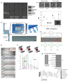
Microfluidic-based measurement techniques. (a) Hydrodynamic stretching by a sudden expansion. Figure adapted from [78] with permission. (b) Deformability index study for RBC parachute shape. Figure adapted from [74] with permission. (c) Constriction method of array design. Figure adapted from [133] with permission. (d) Constriction method of impedance sensing. Figure adapted from [134] under Creative Commons CC BY license. (e). Motion analyses. Figure adapted from [135] with permission. (f) Shape analysis. Figure adapted from [136] with permission. (g). Image and machine learning technique. Figure adapted from [137] under Creative Commons Attribution-Non-commercial-NoDerivatives License 4.0 (CC BY-NC-ND).

References

    1. Smith M.M., Renew J.R., Nelson J.A., Barbara D.W. Red Blood Cell Disorders: Perioperative Considerations for Patients Undergoing Cardiac Surgery. J. Cardiothorac. Vasc. Anesth. 2019;33:1393–1406. doi: 10.1053/j.jvca.2018.08.001. - DOI - PubMed
    1. Kim Y., Kim K., Park Y. Blood Cell—An Overview of Studies in Hematology. IntechOpen; London, UK: 2012. - DOI
    1. Rand R.P., Burton A.C. Mechanical Properties of the Red Cell Membrane I. Membrane Stiffness and Intracellular Pressure. Biophys. J. 1964;4:115–135. doi: 10.1016/S0006-3495(64)86773-4. - DOI - PMC - PubMed
    1. Mohandas N., Clark M.R., Jacobs M.S., Shohet S.B. Analysis of Factors Regulating Erythrocyte Deformability. J. Clin. Investig. 1980;66:563–573. doi: 10.1172/JCI109888. - DOI - PMC - PubMed
    1. An X., Mohandas N. Disorders of Red Cell Membrane. Br. J. Haematol. 2008;141:367–375. doi: 10.1111/j.1365-2141.2008.07091.x. - DOI - PubMed

LinkOut - more resources