. 2025 Sep 11;14(18):1422.
doi: 10.3390/cells14181422.

PA2G4 Functions as a Cofactor for MYC Family Oncoproteins in MYC-Driven Malignancies

Affiliations

PA2G4 Functions as a Cofactor for MYC Family Oncoproteins in MYC-Driven Malignancies

Sukriti Krishan et al. Cells. .

Abstract

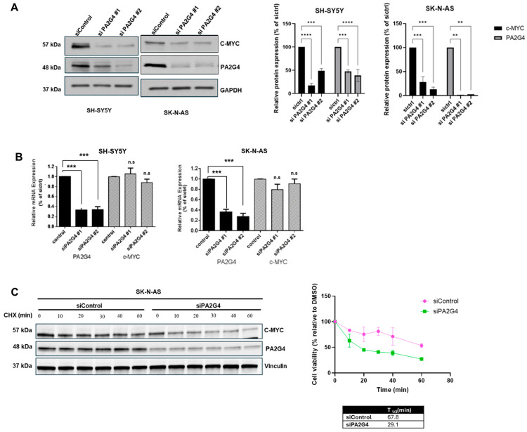
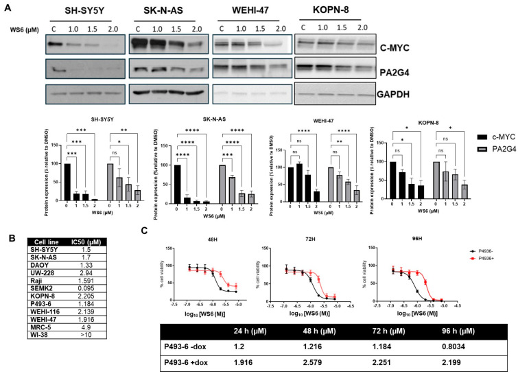
MYCN and c-MYC are critical driver oncogenes in several childhood cancers, including neuroblastoma. Currently, the clinical development of MYC inhibitors has been hindered by the intrinsically disordered structure of MYC proteins, which lack well-defined ligand-binding pockets. Proliferation-associated protein 2G4 (PA2G4) directly binds to and stabilizes MYCN protein, leading to markedly increased MYCN levels in neuroblastoma cells. Here, we demonstrate that PA2G4 is essential for MYCN-driven tumor growth in neuroblastoma in vivo. Moreover, PA2G4 elevates c-MYC protein levels in neuroblastoma cells by inhibiting its ubiquitin-mediated degradation. In turn, c-MYC upregulates the transcription and protein expression of PA2G4, creating an oncogenic feed-forward expression loop. A small molecule PA2G4 inhibitor, WS6, directly disrupts the PA2G4-c-MYC protein-protein interaction, resulting in decreased levels of both PA2G4 and c-MYC. WS6 exhibited selective cytotoxicity in c-MYC-overexpressing cell lines. Together, these findings identify PA2G4 as a shared cofactor for both the c-MYC and MYCN oncoproteins and highlight its interaction with MYC family oncoproteins as a promising therapeutic vulnerability in MYC-driven cancers.

Keywords: Burkitt’s lymphoma; MYC; MYCN; PA2G4; WS6; inhibitors; neuroblastoma.


Conflict of interest statement

The authors declare no conflicts of interest.

Figures

Figure 1
Figure 1
PA2G4 gene knockout decreases neuroblastoma tumorigenesis in Th-MYCN transgenic mice. (A) PA2G4 whole-body knockout mice were generated by deleting exons 1 to 5 of the PA2G4 gene (located on mouse chromosome 10) using a double-stranded DNA break within the PA2G4 locus; (B) Hemizygous PA2G4WT/DelEx1–5 mice were backcrossed with Hemizygous Th-MYCN+/− mice for three generations. (C) Kaplan–Meir Plot depicting proportion of hemizygous Th-MYCN+/− (left panel) or Th-MYCN+/+ (right panel), crossed with either PA2G DelEx1-/DelEx1–5 PA2G4WT/DelEx1–5 or PA2G4WT/WT, surviving tumor-free.
Figure 2
Figure 2
c-MYC regulates PA2G4 expression. (A) Western blot analysis showing PA2G4 and c-MYC protein expression levels in a panel of human neuroblastoma (SH-SY5Y, SK-N-AS), medulloblastoma (DAOY, UW-228), Burkitt lymphoma (Raji and P493-6), acute lymphoblastic leukemia (KOPN-8 and SEM-K2) and Murine B-cell ALL (WEHI-47 and WEHI-116) cell lines, and normal fibroblasts (MRC-5, WI-38) for a correlation between PA2G4 and c-MYC expression in the Western blot (r = 0.57) and CCLE + HPA transcript expression dataset (r = 0.53); (B) Western blot analysis showing PA2G4 and c-MYC protein expression levels after siRNA knockdown of c-MYC in SH-SY5Y cells and SK-N-AS cells at 48 h in comparison to siRNA control and using GAPDH as loading control; (C) Western blot analysis showing PA2G4 and c-MYC protein expression levels after Dox-inducible knockdown of c-MYC in P493-6 cells; (D) real-time PCR analysis of c-MYC and PA2G4 mRNA expression following c-MYC siRNA knockdown in SH-SY5Y and SK-N-AS human neuroblastoma cells. Statistical significance was determined using Student’s t-test. * p < 0.05, ** p < 0.01, *** p < 0.001, **** p < 0.0001, ns not significant.
Figure 3
Figure 3
PA2G4 increases c-MYC protein stability. (A) Immunoblots showing PA2G4 and c-MYC protein expression levels after siRNA knockdown of PA2G4 in SH-SY5Y cells and SK-N-AS cells at 48 h and 96 h, respectively, compared to siControl. GAPDH is a loading control; (B) real-time PCR analysis of c-MYC and PA2G4 mRNA expression following PA2G4 siRNA knockdown in SH-SY5Y and SK-N-AS neuroblastoma cells; (C) knockdown with PA2G4 siRNA in SK-N-AS cells for 48 h, followed by treatment with 100 μg/μL cycloheximide for up to 60 min. Densitometry of c-MYC and PA2G4 protein levels on immunoblotting was used to estimate c-MYC protein half-life. Statistical significance was determined using Student’s t-test. ** p < 0.01, *** p < 0.001, **** p < 0.0001, n.s not significant.
Figure 4
Figure 4
The growth inhibitory effects of WS6 on c-MYC-driven cancer cells. (A) Protein expression levels of PA2G4, c-MYC and loading control, GAPDH, in neuroblastoma (SH-SY5Y, SK-N-AS), B Lymphoma or ALL (WEHI47, KOPN-8) cancer cell lines treated with WS6, a small molecule inhibitor of PA2G4, for 72 h; (B) IC50 concentrations for WS6 in a panel of c-MYC-expressing cancer cell lines and normal fibroblasts after 72 h treatment; (C) cell viability and IC50 values of a c-MYC expressing murine lymphoma cell line with Doxycycline-regulable c-MYC knockdown (P493-6-dox: with c-MYC; and P493-6+dox: without c-MYC) following 48, 72, and 96 h of WS6 treatment (0–8 μM). Statistical significance was determined using Student’s t-test. * p < 0.05, ** p < 0.01, *** p < 0.001, **** p < 0.0001, ns not significant.
Figure 5
Figure 5
Combination therapy for neuroblastoma cells with WS6 and chemotherapeutic agents. Histogram depicting cell viability of SH-SY5Y cells treated with DMSO, WS6, Vincristine, Cisplatin, Etoposide, or WS6 + Vincristine, Cisplatin or Etoposide. Statistical significance was determined using Student’s t-test. * p < 0.05, ** p < 0.01, *** p < 0.001, **** p < 0.0001.

References

    1. Kalkat M., De Melo J., Hickman K.A., Lourenco C., Redel C., Resetca D., Tamachi A., Tu W.B., Penn L.Z. MYC Deregulation in Primary Human Cancers. Genes. 2017;8:151. doi: 10.3390/genes8060151. - DOI - PMC - PubMed
    1. Schick M., Habringer S., Nilsson J.A., Keller U. Pathogenesis and therapeutic targeting of aberrant MYC expression in haematological cancers. Br. J. Haematol. 2017;179:724–738. doi: 10.1111/bjh.14917. - DOI - PubMed
    1. Chen X., Bahrami A., Pappo A., Easton J., Dalton J., Hedlund E., Ellison D., Shurtleff S., Wu G., Wei L. Recurrent somatic structural variations contribute to tumorigenesis in pediatric osteosarcoma. Cell Rep. 2014;7:104–112. doi: 10.1016/j.celrep.2014.03.003. - DOI - PMC - PubMed
    1. Edmunds L.R., Sharma L., Kang A., Lu J., Vockley J., Basu S., Uppala R., Goetzman E.S., Beck M.E., Scott D., et al. c-Myc programs fatty acid metabolism and dictates acetyl-CoA abundance and fate. J. Biol. Chem. 2015;290:20100. doi: 10.1074/jbc.A114.580662. - DOI - PMC - PubMed
    1. Chou Y.T., Lin H.H., Lien Y.C., Wang Y.H., Hong C.F., Kao Y.R., Lin S.C., Chang Y.C., Lin S.Y., Chen S.J., et al. EGFR promotes lung tumorigenesis by activating miR-7 through a Ras/ERK/Myc pathway that targets the Ets2 transcriptional repressor ERF. Cancer Res. 2010;70:8822–8831. doi: 10.1158/0008-5472.CAN-10-0638. - DOI - PubMed

Substances

LinkOut - more resources