. 2025 Sep 26;15(1):124.
doi: 10.1186/s13550-025-01283-x.

18F-BMS-986229 PET imaging of tumor PD-L1 expression in glioblastoma patients

Affiliations

18F-BMS-986229 PET imaging of tumor PD-L1 expression in glioblastoma patients

Milan Grkovski et al. EJNMMI Res. .

Abstract

Background: Neurooncologists urgently need biomarkers that can optimize the clinical development of PD-L1 targeted immunotherapy in the treatment of glioblastoma. This study evaluated PD-L1 targeted tumor imaging by positron emission tomography (PET) in glioblastoma patients, using the novel macrocyclic peptide radiotracer 18F-BMS-986229.

Results: Twelve adult postsurgical glioblastoma patients underwent brain PET imaging 1-hour post injection 190±20 MBq of 18F-BMS-986229. In a subset of patients, dynamic PET scans were obtained for pharmacokinetic modeling in tumors and normal tissues. Tracer kinetics in both tumor sites and normal tissues were well described by a reversible 1-tissue compartment model. Tumor sites demonstrated 18F-BMS-986229 tracer-avidity (SUV = 1.1 ± 0.4; range, 0.6-1.7) in 10 of 12 cases, with negligible tracer-avidity in normal brain structures. Tumor avidity for 18F-BMS-986229 on PET was spatially independent of tumor contrast-enhancement on magnetic resonance imaging, indicating that tracer-binding at tumor site was not dependent upon blood-brain barrier breakdown. The observed tumor site low tracer-uptake paralleled low immunohistochemical PD-L1 expression in resected tumors, with no correlations between standardized uptake value versus tumor MGMT methylation, PTEN oncogenic mutation status, tumor mutation burden, or patient overall survival.

Conclusion: This pilot study demonstrates the feasibility of characterizing tumor sites in glioblastoma patients by PD-L1-targeted PET imaging with18F-BMS-986229, even in patients with low tumor PD-L1 expression. We hypothesize that 18F-BMS-986229 PET can improve the pharmacometrics of PD-L1-targeted therapy trials.

Trial registration number: NCT02617589. Trial Registration Date: December 1st, 2015.

Keywords: 18F-BMS-986229; Glioblastoma; PD-L1 expression; Pharmacokinetic modeling.


Conflict of interest statement

Declarations. Ethics approval and consent to participate: This study was approved by the Institutional Review Board (Protocol #16–078, ClinicalTrials.gov Identifier: NCT02617589) and performed under an investigational new drug application approved by U.S. Food and Drug Administration. The analysis was conducted following the principles outlined in the Declaration of Helsinki. Patient clinical information was collected anonymously from electronic medical records. Consent for publication: All patients provided written informed consent. Competing interests: IKM reports receiving personal fees (advisory board services) from Agios, Black Diamond Therapeutics, Debiopharm Group, Erasca, Novartis, Prelude Therapeutics, Roche Therapeutics, Servier Pharmaceuticals, Voyager. IKM reports receiving grants from General Electric and Puma Biotechnology. No potential conflicts of interest relevant to this article exist.

Figures

Fig. 1
Fig. 1
68 year old male [Patient #1]. MRI (left) shows blood products within resection cavity and enhancing mural nodularity consistent with tumor which demonstrates avidity for 18 F-BMS-986229. SUV display intensity scale (0.0-2.5). H&E sections (right) demonstrate malignant astrocytic cells with readily identifiable mitoses, consistent with glioblastoma. Scale bar (bottom left) = 100 μm
Fig. 2
Fig. 2
72 year old female [Patient #8]. 18 F-BMS-986229 PET/MRI fusion (left) of a single axial plane focusing on tumor in the left cerebral hemisphere. MRI demonstrates uniform contrast-enhancement around surgical cavity. Nodular tissue enhancement along anterior margin of the cavity demonstrates prominent tracer-avidity, with minimal avidity in other enhancing tissues. Contrast-enhancing inflammation around craniotomy showed tracer-avidity. SUV display intensity scale (0.0-2.5). H&E sections (middle) demonstrate malignant astrocytic cells with readily identifiable mitoses, consistent with glioblastoma. Membranous labeling of tumor cells by PDL1 immunohistochemical stains was 0% (negative; right). Scale bar (bottom left) = 100 μm
Fig. 3
Fig. 3
57 year old F [Patient #2]. 18 F BMS-986229 PET/MRI fusion (top left) of a single axial plane showing tumor in right cerebral hemisphere. MRI demonstrates uniform contrast-enhancement in tumor. Lateral subregion of enhancing tumor demonstrates prominent tracer-avidity, with minimal avidity in remainder of enhancing tumor. SUV display intensity scale (0.0-2.5). Time-activity curves for structures of interest (top right). 18 F-BMS-986229 SUV as a function of imaging time post-injection for small intratumor area with higher uptake and for normal structures. Measured data (circles) are presented with fits from a 1-tissue reversible compartment model. H&E sections (bottom left) demonstrate malignant astrocytic cells with readily identifiable mitoses, consistent with glioblastoma. Membranous labeling of tumor cells by PDL1 immunohistochemical stains was 0% (negative; bottom right). Scale bar (bottom left) = 100 μm
Fig. 4
Fig. 4
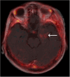
69 year old female [Patient #10] with GBM status post resection with recurrent tumor (white arrow). 18 F BMS-986229 PET/MRI fusion. Radiotracer-avid tumor was non-enhancing on MRI. SUV display intensity scale (0.0-2.5)

References

    1. Iivanainen S, Koivunen JP. Possibilities of improving the clinical value of immune checkpoint inhibitor therapies in Cancer care by optimizing patient selection. Int J Mol Sci. 2020;21. - PMC - PubMed
    1. Tamura R, Tanaka T, Morimoto Y, et al. Alterations of the tumor microenvironment in glioblastoma following radiation and Temozolomide with or without bevacizumab. Ann Transl Med. 2020;8:297. - PMC - PubMed
    1. Bensch F, van der Veen EL, Lub-de Hooge MN, et al. (89)Zr-atezolizumab imaging as a non-invasive approach to assess clinical response to PD-L1 Blockade in cancer. Nat Med. 2018;24:1852–8. - PubMed
    1. Restrepo P, Yong R, Laface I et al. Tumoral and immune heterogeneity in an anti-PD-1-responsive glioblastoma: a case study. Cold Spring Harb Mol Case Stud. 2020;6. - PMC - PubMed
    1. Munari E, Zamboni G, Lunardi G, et al. PD-L1 expression heterogeneity in Non-Small cell lung cancer: defining criteria for harmonization between biopsy specimens and whole sections. J Thorac Oncol. 2018;13:1113–20. - PubMed

Associated data

LinkOut - more resources