. 2025 Sep 26;16(1):8480.
doi: 10.1038/s41467-025-63343-z.

Circular RNA-based protein replacement therapy mitigates osteoarthritis in male mice

Affiliations

Circular RNA-based protein replacement therapy mitigates osteoarthritis in male mice

Jinlong Suo et al. Nat Commun. .

Abstract

In vitro-transcribed and circularized RNAs (ivcRNAs) represent a robust platform for sustained protein translation, offering promising potential for localized therapeutic delivery in joint diseases. Osteoarthritis (OA), the most prevalent degenerative joint disorder, remains a major clinical challenge due to its progressive nature and the lack of disease-modifying treatments. In this study, we identify Musashi2 (Msi2) deficiency in articular chondrocytes as a key contributor to OA pathogenesis. To evaluate the efficacy of ivcRNA-mediated protein replacement therapy, we developed a localized delivery strategy that enables high-yield and prolonged protein expression in chondrocytes. Using a destabilization of the medial meniscus (DMM) mouse model, we demonstrate that intra-articular delivery of ivcRNA encoding MSI2 effectively mitigates OA progression in male mice. Furthermore, therapeutic supplementation of SOX5, a downstream effector of MSI2, via ivcRNA delivery further validates this approach. Our findings establish ivcRNA-based protein replacement as a potential RNA therapeutic strategy for osteoarthritis.


Conflict of interest statement

Competing interests: W.Z., L.C., J.S., and L.L. are named as inventors on patents related to circRNA held by CAS CEMCS. L.C. is a scientific co-founder of RiboX Therapeutics. The remaining authors declare no competing interests.

Figures
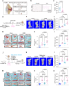
Fig. 1
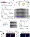
Fig. 1. MSI2 is a potential target for osteoarthritis treatment.
a Schematic illustration of MSI2 function in three experimental models, including aging-induced osteoarthritis (OA) model, DMM-induced OA model, and human OA cartilage. DMM, destabilization of the medial meniscus. b Immunohistochemical staining of MSI2 was performed on paraffin sections of human cartilage from non-weight bearing areas (Control) and weight bearing areas (OA) in patients after arthroplasty. Scale bars = 500 μm. c Quantitative analysis of immunohistochemical staining of MSI2 using Image J software (Each group consists of n = 15 samples). The data are presented as the Mean with SEM. Statistical significance was determined by two-tailed unpaired t test. d Experimental design of tamoxifen (TAM) injection and DMM surgery in Msi2 CKO mice and littermate controls. e Micro-CT scanning showing calcified meniscus and extra bone in joints from Msi2 CKO mice after DMM surgery (calcified meniscus and extra bone are marked in red) (n = 7 mice per group). f Quantification of the bone volume (BV) of the calcified meniscus and extra bone in (e) (n = 7 mice per group). The data are presented as the Mean with SEM. Statistical significance was determined by Ordinary one-way ANOVA. g Representative images by SO&FG staining of paraffin sections from Msi2 CKO mice subjected to the DMM model 8 weeks later (n = 7 mice per group). Scale bars=500μm (top), 200μm (bottom). h Osteoarthritis Research Society International (OARSI) histopathological score of SO&FG staining in (g) (n = 7 mice per group). The data are presented as the Mean with SEM. Statistical significance was determined by Ordinary one-way ANOVA. i Quantification of subchondral bone plate (SBP) thickness in (g) (n = 7 mice per group). The data are presented as the Mean with SEM. Statistical significance was determined by Ordinary one-way ANOVA. j Schematic of intra-articular injection of AAV expressing MSI2 in DMM-treated mice to evaluate the therapeutic effects against OA. AAV-EGFP shown as the negative control, AAV-Msi2RBDmut shown as mutation of MSI2 RNA binding domain. k Micro-CT scanning showing calcified meniscus and extra bone in joints from DMM-treated mice after intra-articular injection of AAV-EGFP, -MSI2, -Msi2RBDmut (calcified meniscus and extra bone are marked in red) (n = 11,10,10,11 mice). l Quantification of the BV of the calcified meniscus and extra bone (n = 11,10,10,11 mice) in k. The data are presented as the Mean with SEM. Statistical significance was determined by Ordinary one-way ANOVA. m Representative images of SO&FG staining from DMM-treated mice after intra-articular injection of AAV-EGFP, -MSI2, -Msi2RBDmut (n = 10 mice per group). Scale bars=500μm (top), scale bars=200 μm (bottom). n OARSI histopathological score of SO&FG staining in (m) (n = 10 mice per group). The data are presented as the Mean with SEM. Statistical significance was determined by Ordinary one-way ANOVA. o Quantification of SBP thickness in (m) (n = 10 mice per group). The data are presented as the Mean with SEM. Statistical significance was determined by Ordinary one-way ANOVA. Figure 1a, d, j: Created in BioRender. Suo, J. (2025) https://BioRender.com/4byajfm.
Fig. 2
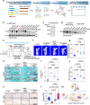
Fig. 2. Engineered circRNAs encoding fLuc exhibit stable and efficient expression in chondrocytes in vivo.
a Schematic diagram of ivcRNA design and synthesis in vitro based on permuted Anabaena group I intron optimized with truncated extraneous sequences (Ana_PIE_27nt system). ivcRNA, in vitro transcribed and circularized RNA. b Schematic diagram of ivcRNA-LNPs (lipid nanoparticles) formulation. The lipid mixture in ethanol and circRNA in aqueous solution were pumped separately into the two inlets of the microfluidic mixing device with a total flow rate of 4 mL/min. c Representative image of ivc-IRES3-fLuc is analyzed using 4% urea denaturing polyacrylamide gel electrophoresis (Urea-PAGE). Bans of RNA circles are enriched and verified with RNase R. The hollow blue circles indicate circular RNAs, single blue curves indicate nicked RNAs. d The pipeline for administering mice to characterize the expression of mRNA/circRNA encoding fLuc in vivo. Intra-articular injection of DiR labeled RNA-LNPs complexes was performed on 6-week-old C57/B6 mice (300 ng RNA per mouse). Bioluminescence imaging of fLuc activity was conducted until the signal returned to background levels. DiR, 1,1’-Dioctadecyl-3,3,3’,3’-Tetramethylindotricarbocyanine Iodide; PBS, phosphate-buffered saline. e Represent bioluminescence IVIS images at various time points of the live mice after intra-articular injection of DiR labeled RNA-LNPs complexes. IVIS, in vivo imaging system spectrum. f Quantitative statistical analysis of the fluorescence at the injection site for in vivo Luciferase expression at various time points (n  =  3 mice per group). The data are presented as the Mean with SD. Statistical significance was determined by multiple two-tailed unpaired t tests in fLuc mRNA group and ivc-IRES3-fLuc group. g Schematic illustration of the experimental pipeline characterizes circRNA injection in sham and DMM-treated mice. Sham (Left leg) and DMM surgery (right leg) were performed in 8-week-old wild-type mice. The dosage of circRNA injection is 500 ng. h Bioluminescent images of a representative mice. n = 4 mice per group. Left leg: Sham group; right leg: DMM group. i Quantitative statistical analysis of the fluorescence at the injection site (Sham and DMM) for in vivo Luciferase expression (n  =  4 mice per group). The data are presented as the violin plot and the number of mice is showed as points in each group. Statistical significance was determined by multiple two-tailed unpaired t test, and p-values are labelled in (i). j Representative image of ivc-IRES4-mCherry RNA is analyzed by 4% urea-PAGE. RNA circles are enriched and verified with RNase R. The hollow red circles indicate RNA circles, single red curves indicate nicked RNA. k Immunofluorescence staining of mCherry in articular cartilage of joints from mice after intra-articular injection of ivc-IRES4-mCherry-LNPs. n = 3 samples per group. Scale bars = 100 µm. l Representative image of ivc-IRES2-EGFP is analyzed using denaturing 4% urea-PAGE. RNA circles are enriched and verified with RNase R. The hollow green circles indicate RNA circles; single green curves indicate nicked RNA. m Representative microscopy immunofluorescence staining images of a cross section of day-3 cartilage pellet cultures after incubated with ivc-IRES2-EGFP-LNPs. n = 3 samples per group. Scale bars = 100 µm. Figure 2 a, b, d, g. k, m: Created in BioRender. Suo, J. (2025) https://BioRender.com/4byajfm.
Fig. 3
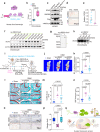
Fig. 3. ivcRNA-based MSI2 therapy alleviates osteoarthritis.
a Schematic showing the pipeline of designing and screening engineered ivcRNA encoding MSI2. First, we select different types of IRES elements screened from the viral IRES database, then, optimize the coding sequence (cargo) and combine various IRES elements with the cargo sequence, finally screen combinations of IRES-cargo to obtain IRES functional folding. The prepared in vitro transcribed circRNA deliver into chondrocyte cells to obtain the best ivcRNA of IRES-cargo combination using western blotting (WB). b WB analysis of mouse MSI2 expression levels in chondrocytes 24 h after transfection with various IRES-Msi2 combinations. ACTIN was used as a reference protein. Quantification of MSI2 bands was performed with Image J software. The IRES-Msi2-Opt1 and IRES-Msi2-Opt2 plasmids contain a 3 × Flag tag, while the IRES-Msi2-Opt3 plasmids do not. Opt: Optimized. c Representative image of ivc-IRES4-Msi2 is analyzed using 4% denaturing urea-PAGE. RNA circles are enriched and verified with RNase R treatment. The hollow violet circles indicate RNA circles; single violet curves indicate nicked RNAs. d WB analysis of mouse MSI2 expression levels in chondrocytes after transfection with ivc-IRES4-Msi2-3×Flag and Msi2 mRNA. Quantification of MSI2 bands was performed with Image J software. e Schematic illustration of intra-articular injection of ivc-IRES4-Msi2-LNPs to treatment DMM mice. Two group, Sham and DMM mice, were treated at 10-week-old and monitored to evaluate the therapeutics effects against OA caused by DMM surgery after 8-week-teatment. f Micro-CT scans show calcified meniscus and extra bone after intra-articular injection of ivc-IRES3-fLuc, Msi2 mRNA and ivc-IRES4-Msi2 in DMM model mice (calcified meniscus and extra bone are marked in red) (n = 12, 10, 10, 10 mice). g Quantification of the BV of the calcified meniscus and extra bone in (f) (n = 12, 10, 10, 10 mice). The data are presented as the Mean with SEM. Statistical significance was determined by Ordinary one-way ANOVA. h Representative images of SO&FG staining of paraffin sections from wild-type mice injected with linear and ivcRNA encoding MSI2 and ivcRNA encoding fLuc after DMM (n = 9 mice per group). Scale bars=100μm. i OARSI histopathological score for SO&FG staining in (h) (n = 9 mice per group). The data are presented as the Mean with SEM. Statistical significance was determined by Ordinary one-way ANOVA. j Quantification of SBP thickness in (h) (n = 9 mice per group). The data are presented as the Mean with SEM. Statistical significance was determined by Ordinary one-way ANOVA. k Immunohistochemical staining with MSI2 antibody was performed on paraffin sections of knee joints from wild-type mice injected with ivc-IRES3-fLuc, Msi2 mRNA and ivc-IRES4-Msi2 after DMM surgery (n = 5 samples per group), scale bars = 200 μm (top), scale bars = 100 μm (bottom). l Quantification of MSI2 signals in (k) by Image J software (n = 5 samples per group). The data are presented as the box-and-whisker plots show minimum (Min) to maximum (Max) and all points. Statistical significance was determined by Ordinary one-way ANOVA. Boxes: 25th–75th percentiles; centre line: median; whiskers: 1.5 × IQR. m Schematic representation of ivcRNA-based therapy compensates MSI2 to alleviate the phenotypes of OA in DMM mice. The experiments in Fig. 3b, c, d was repeated three times independently and similar results were obtained. The images of knee joint, RNA and mice in Fig. 3a, e, m were adapted from BioRender.com. Created in BioRender. Suo, J. (2025) https://BioRender.com/4byajfm.
Fig. 4
Fig. 4. ivcRNA-based SOX5 therapy inhibits osteoarthritis progression.
a Schematic illustration of the mouse Sox5 transcript (ENSMUST00000038815.13) binding by MSI2 protein. Blue Bars, the putative MSI2 binding elements (MBEs: r(G/A)U1–3AGU). CDS, coding sequence. b Coimmunoprecipitated RNAs were analyzed for the enrichment of Sox5 transcripts with anti-MSI2 antibody or a control rabbit IgG in chondrocytes. The data are presented as the box-and-whisker plots show Min to Max and all points. n = 4 biological repeats. Statistical significance was determined by two-tailed unpaired t test. c WB analysis of SOX5, HMGB2, VINCULIN and MSI2 protein levels in ATDC5 cells overexpressing Flag-tagged Msi2 and Flag-tagged Msi2RBDmut. VINCULIN was used as a reference protein. d Immunohistochemical staining with SOX5 antibody was performed on paraffin sections of cartilage from patients with arthroplasty. Scale bars = 200 μm. e Quantitative statistics of signals from immunohistochemical staining of SOX5 in (d) using Image J software (n = 9 samples per group). The data are presented as the Mean with SEM. Statistical significance was determined by two-tailed unpaired t test. f WB analysis of mouse SOX5 expression levels in chondrocyte after 24 h of transfection with various IRES-Sox5 combinations and Sox5 mRNA. ACTIN was used as a reference protein. Quantification of SOX5 bands was performed with Image J software. g Representative image of ivc-IRES3-Sox5 is analyzed using 4% denaturing urea-PAGE. RNA circles are enriched and verified with RNase R treatment. The hollow green circles indicate RNA circles; single green curves indicate nicked RNAs. h WB analysis of mouse SOX5 expression levels in chondrocytes after transfection with ivc-IRES3-Sox5. Quantification of SOX5 bands was performed with Image J software. i Schematic illustration of intra-articular injection of ivcRNA-LNPs encoding SOX5 to treatment DMM mice. ivcRNA-based therapy treated 10-week-old mice of sham and DMM. Then, the therapeutic phenotypes of OA were evaluated in sham and DMM mice after 7-week-treatment by micro-CT, SO&FG staining and immunohistochemistry. j Micro-CT scans show calcified meniscus and extra bone after intra-articular injection of ivc-IRES3-fLuc-LNPs, and ivc-IRES3-Sox5-LNPs in DMM model mice (calcified meniscus and extra bone are marked in red) (n = 10, 10, 20 mice). k Quantification of the BV of the calcified meniscus and extra bone in (j) (n = 10, 10, 20 mice). The data are presented as the Mean with SEM. Statistical significance was determined by Ordinary one-way ANOVA. l Representative images of SO&FG staining of paraffin sections from wild-type mice injected with ivcRNA encoding SOX5 and ivcRNA encoding fLuc after DMM surgery (n = 10, 10, 19 mice). Scale bars = 100 μm. m OARSI histopathological score for SO&FG staining in (l) (n = 10, 10, 19 mice). The data are presented as the Mean with SEM. Statistical significance was determined by Ordinary one-way ANOVA. n Quantification of SBP thickness in (l) (n = 10, 10, 19 mice). The data are presented as the Mean with SEM. Statistical significance was determined by Ordinary one-way ANOVA. o Immunohistochemical staining with SOX5 antibody was performed on paraffin sections of knee joint from wild-type mice injected with ivcRNA encoding SOX5 and ivcRNA encoding fLuc after DMM surgery (n = 6 samples per group). Scale bars = 200 μm (top), scale bars = 50 μm (bottom). p Quantitative statistics of signals from immunohistochemical staining of SOX5 in (o) using Image J software (n = 6 samples per group). The data are presented as the box-and-whisker plots show Min to Max and all points. Statistical significance was determined by Ordinary one-way ANOVA. q Schematic representation of ivcRNA-based therapy compensates SOX5, a downstream target of MSI2, to alleviate the phenotypes of OA in DMM mice. MSI2 can bind to the 3’UTR of Sox5 transcripts to promote SOX5 translation, and DMM surgery resulted in a significant decrease in SOX5 protein expression. Injecting ivcRNA-LNPs into the joint cavity of mice after DMM surgery to effectively express SOX5 protein in chondrocytes. Figure 4b, p: Boxes: 25th–75th percentiles; centre line: median; whiskers: 1.5× IQR. The experiments in Fig. 4c, f, g, h was repeated three times independently and similar results were obtained. Figure 4i, q:Created in BioRender. Suo, J. (2025) https://BioRender.com/4byajfm.

References

    1. Baden, L. R. et al. Efficacy and Safety of the mRNA-1273 SARS-CoV-2 Vaccine. N. Engl. J. Med384, 403–416 (2021). - PMC - PubMed
    1. Walsh, E. E. et al. Safety and immunogenicity of two RNA-based Covid-19 vaccine candidates. N. Engl. J. Med383, 2439–2450 (2020). - PMC - PubMed
    1. Parhiz, H., Atochina-Vasserman, E. N. & Weissman, D. mRNA-based therapeutics: looking beyond COVID-19 vaccines. Lancet403, 1192–1204 (2024). - PubMed
    1. Qin, S. et al. mRNA-based therapeutics: powerful and versatile tools to combat diseases. Signal Transduct. Target Ther.7, 166 (2022). - PMC - PubMed
    1. Rohner, E., Yang, R., Foo, K. S., Goedel, A. & Chien, K. R. Unlocking the promise of mRNA therapeutics. Nat. Biotechnol.40, 1586–1600 (2022). - PubMed

LinkOut - more resources