. 2025 Sep 26;15(1):33168.
doi: 10.1038/s41598-025-13997-y.

Development of a novel assay for antigen presentation measurement

Affiliations

Development of a novel assay for antigen presentation measurement

Mei Li et al. Sci Rep. .

Abstract

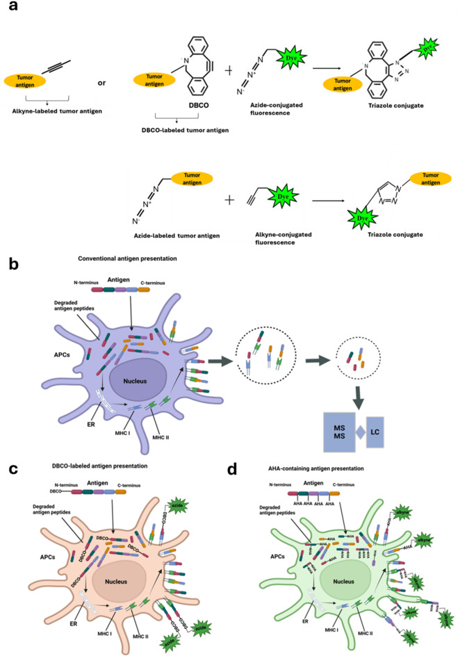
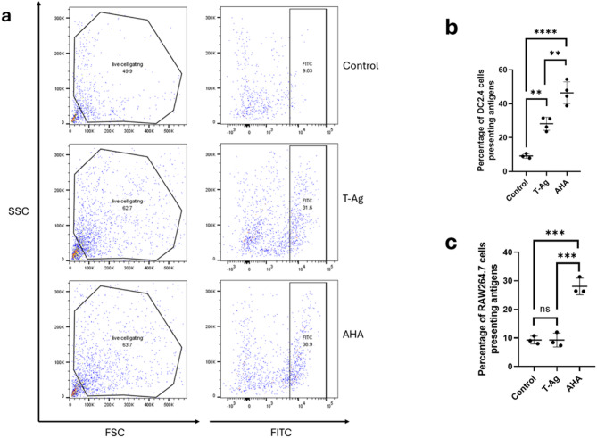
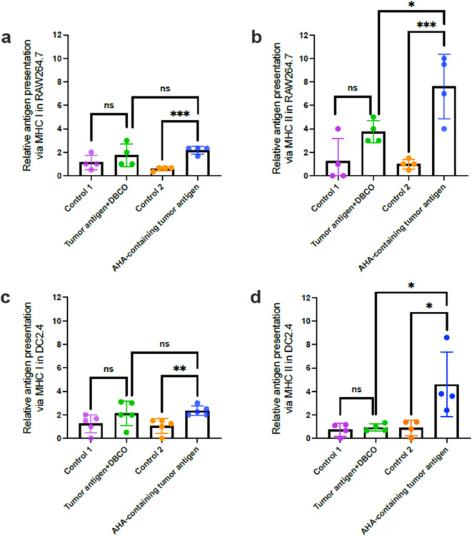
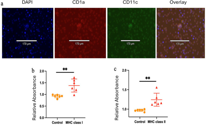
Accurate measurement of antigen presentation is essential for understanding immune responses to infections and tumors. However, current methods are cumbersome, time-consuming, and rely on known peptide sequences and antibodies, leading to unstable antigen presentation, antigen loss during processing and editing, and inconsistent results. We developed a novel, cost-effective method for examining antigen presentation using Click chemistry, which utilizes a bioorthogonal reaction between azides and alkynes/cyclooctenes. Antigens were pre-labeled with azides or alkynes to facilitate their uptake by antigen-presenting cells (APCs). Their presentation was subsequently detected using fluorophore-conjugated dibenzocyclooctyne or azide. The study involved three types of APCs, mouse macrophages (RAW264.7), mouse dendritic cells (DC2.4), and mouse primary bone marrow derived dendritic cells (BMDCs), and three categories of antigens: BSA, bacteria, and tumor antigens. Antigen presentation was measured and validated through multiple analytic techniques, including a fluorescent plate reader, flow cytometry, and ELISA. We showed efficient and stable presentation of antigens on the surface of all RAW264.7, DC2.4 and BMDCs. Antigens labeled using Click chemistry showed enhanced stability within the phagolysosomes of APCs. Notably, antigens labeled throughout the peptide sequence using azidohomoalanine (AHA) exhibited superior presentation on MHC class II compared to antigens labeled only at the N-terminus. Furthermore, this method preserved the natural antigen editing process, enabling the selection of high-affinity antigens for MHC presentation. This novel antigen presentation assay offers key advantages over existing methods, including faster processing, cost-effectiveness, stable antigen presentation, and reliable detection signals. When paired with mass spectrometry, it can identify stably presented tumor peptides, offering potential targets for immunotherapy development.

Keywords: Antigen presentation; Breast cancer; Click chemistry; MHC I/II; Phagocytosis.


Conflict of interest statement

Declarations. Competing interests: The authors declare no competing interests.

Figures

Fig. 1
Fig. 1
Chemical scheme outlining the click-chemistry use in this study and comparison of conventional and our novel antigen presentation assays. Click-chemistry is a reaction between azides and alkynes/cyclooctenes. Thus, tumor antigens labeled with DBCO or azide can be detected using fluorescent probes conjugated with azide or alkyne, respectively (a). The stability and reliability of antigen presentation in conventional assays heavily depend on the sequence of the predefined antigen (b). Terminal labeling with DBCO across diverse antigens enables APCs to select the antigen(s) that best fit MHC molecules but introduces the risk of N-terminal truncation during antigen processing (c). In contrast, integrating AHA throughout the entire antigen across diverse antigens increases the likelihood that click signals remain detectable in the final presented antigens, regardless of how the antigens are processed and selected (d).
Fig.2
Fig.2
Azido- and alkyne-labeled amino acids demonstrate stability within phagolysosomes. Triple immunofluorescence staining was performed to evaluate the phagocytosis of DBCO-labeled tumor antigens (a) and AHA-containing tumor antigens (b) by macrophages. Blue fluorescence marks the nucleus, green fluorescence labels the phagolysosome, and red fluorescence identifies the DBCO-labeled or AHA-containing tumor antigens. Macrophages treated without tumor antigens served as controls. The mean fluorescence intensity of the green signal, indicative of phagocytosis, was quantified (c). Data are shown as mean ± SD. (*, p < 0.05). DBCO, dibenzocyclooctyne; AHA, L-Azidohomoalanine.
Fig. 3
Fig. 3
Antigen presentation of DBCO-conjugated antigens by APCs. Mouse macrophages (ac) and mouse dendritic cells (df) were cultured with N-terminally DBCO-labeled bacterial antigens (E. coli), BSA, and tumor antigens for six days. Surface presentation of DBCO-labeled antigens was detected using the membrane-impermeable azide–Alexa Fluor 488 dye. The resulting fluorescence intensity reflected the surface display of DBCO-labeled E. coli, BSA, and tumor antigens. Untreated cells served as controls. Data are shown as mean ± SD. (*, p < 0.05; ***, p < 0.001; ns, no statistical difference).
Fig. 4
Fig. 4
Flow cytometry plots and graphs comparing the surface presentation of fluorescent DBCO- and AHA-labeled tumor antigens by APCs. (a) Representative flow cytometry gating strategy showing antigen presentation in dendritic cells untreated (top), treated with DBCO-labeled tumor antigens (T-Ag, middle), and treated with AHA-containing tumor antigens (AHA, bottom). (bc) Histograms illustrating the effectiveness of antigen presentation by dendritic cells (b) and macrophages (c) for DBCO-labeled and AHA-containing tumor antigens. Data are shown as mean ± SD. (**, p < 0.01; ***, p < 0.001; ****, p < 0.0001; ns, no statistical difference).
Fig. 5
Fig. 5
Extracellular DBCO- and AHA-labeled tumor antigens are predominantly presented on MHC class II, with notable cross-presentation on MHC class I. RAW264.7 (ab) and DC2.4 (cd) cells were cultured with DBCO-labeled or AHA-containing tumor antigens for six days. After fixation and cross-linking, MHC I– or MHC II–presented antigens were captured on plates pre-coated with corresponding antibodies. Antigen presentation was detected via click chemistry using azide- or alkyne-linked HRP, and quantified based on HRP-derived absorbance. Data are presented as mean ± SD. (*, p < 0.05; **, p < 0.01; ***, p < 0.001; ns, no statistical difference).
Fig. 6
Fig. 6
Evaluation of AHA-incorporated tumor antigen presentation via MHC I and MHC II in primary dendritic cells derived from mouse bone marrow. (a) Dendritic cell identity was confirmed using CD1a and CD11c (both known as dendritic cell markers). Antigen presentation of AHA-labeled tumor antigens was assessed using a sandwich ELISA. Significant antigen presentation through MHC class I (b) and MHC class II (c) was determined by comparing absorbance values to control background levels. Data are presented as mean ± SD. (**, p < 0.01, compared to control group).

References

    1. Shi, C. et al. ultifactorial Diseases of the Heart, Kidneys, Lungs, and Liver and Incident Cancer: Epidemiology and Shared Mechanisms. Cancers (Basel)15(3), 729 (2023). - PMC - PubMed
    1. Wishart, D. S. Is cancer a genetic disease or a metabolic disease? EBioMedicine2 (6), 478–479 (2015). - PMC - PubMed
    1. Hartmaier, R. J. et al. Genomic analysis of 63,220 tumors reveals insights into tumor uniqueness and targeted cancer immunotherapy strategies. Genome. Med.9(1), 16 (2017). - PMC - PubMed
    1. Yang, R. & Cui, J. Advances and applications of RNA vaccines in tumor treatment. Mol. Cancer. 23 (1), 226 (2024). - PMC - PubMed
    1. Trivedi, V. et al. mRNA-based precision targeting of neoantigens and tumor-associated antigens in malignant brain tumors. Genome. Med.16(1), 17 (2024). - PMC - PubMed

LinkOut - more resources