. 2025 Sep 2;14(9):1171.
doi: 10.3390/biology14091171.

High Salt Intake Affects Visceral Adipose Tissue Homeostasis: Beneficial Effects of GLP-1 Agonists

Affiliations

High Salt Intake Affects Visceral Adipose Tissue Homeostasis: Beneficial Effects of GLP-1 Agonists

Vanessa Touceda et al. Biology (Basel). .

Abstract

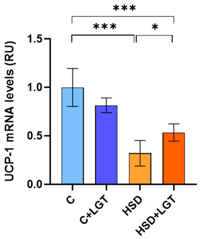
High salt (NaCl) intake has been associated with visceral adipose tissue (VAT) dysfunction independently of its impact on blood pressure. Liraglutide (LGT), a GLP-1 agonist, could be a potential therapeutic option. We investigated the impact of a chronic high-salt diet (HSD) on VAT homeostasis and evaluated the potential protective effects of LGT, a GLP-1 receptor agonist. Male C57BL/6 mice were fed a standard diet (Control, C) or 8% NaCl (HSD) for 15 weeks and subsequently treated with LGT or vehicle for 5 weeks. In VAT, histological characteristics, collagen deposition, vascular density, mitochondrial dynamics, oxidative stress, and adipokine expression were evaluated. The HSD significantly decreased body weight, VAT mass, and adipocyte size (p < 0.05). Moreover, it impaired vascular density and induced interstitial fibrosis (p < 0.01). LGT treatment improved vascularization and VEGF expression and reduced fibrosis (p < 0.05 vs. the HSD). The HSD induced oxidative stress and mitochondrial fragmentation, which were attenuated by LGT (p < 0.001). Leptin levels were elevated by the HSD (p < 0.05) and normalized with LGT, while adiponectin levels increased. In conclusion, excessive salt consumption induces structural and metabolic dysfunction in VAT. LGT therapy mitigates several of these adverse effects, supporting its potential as a novel strategy for managing salt-sensitive adipose tissue dysfunction.

Keywords: adipose tissue; fibrosis; high salt intake; liraglutide; vascular density.


Conflict of interest statement

The authors declare no conflicts of interest.

Figures

Figure 1
Figure 1
Histological characteristics of EAT. (A) Representative images of H-E-stained sections; (B) histological characteristics of EAT in the Control (C), Control + LGT (C + LGT), HSD, and HSD + LGT groups. Data are presented as means ± SDs. One-way ANOVA followed by Bonferroni’s post hoc test. * p < 0.05, ** p < 0.01.
Figure 2
Figure 2
Interstitial collagen content in visceral adipose tissue. (A) Representative images of Picrosirius Red-stained sections. (B) Collagen volume fraction (%) quantification and total collagen content measured through the hydroxyproline assay in the Control (C), Control + LGT (C + LGT), HSD, and HSD + LGT groups. Data are presented as means ± SDs. One-way ANOVA followed by Bonferroni’s post hoc test. * p < 0.05, ** p < 0.01.
Figure 3
Figure 3
Vascular density measured by a fluorescence assay. (A) Representative images of rhodamine-labeled GSL lectin, fluorescein–peanut agglutinin lectin and DAPI–stained sections of representative EAT samples from the Control (A), Control + LGT (B), HSD (C), and HSD + LGT (D) groups. (B) Quantification of vascular density and its correlation with the total collagen content (Spearman’s test, n = 10). Data are presented as means ± SDs. One-way ANOVA followed by Bonferroni’s post hoc test. * p < 0.05.
Figure 4
Figure 4
Immunohistochemical analysis of Vascular Endothelial Growth Factor expression. Representative images of EAT from the Control (C), Control + LGT (C + LGT), HSD, and HSD + LGT groups. VEGF levels are expressed as percentages (%) of the total tissue area. Data are presented as means ± SDs. One-way ANOVA followed by Bonferroni’s post hoc test. * p < 0.05, ** p < 0.01.
Figure 5
Figure 5
HIF-1α expression. HIF-1a mRNA levels in epididymal adipose tissue (EAT) from the Control (C), Control + LGT (C + LGT), HSD, and HSD + LGT groups. Data are presented as means ± SDs. One-way ANOVA followed by Bonferroni’s post hoc test. *** p < 0.001.
Figure 6
Figure 6
Oxidative stress markers. Catalase and superoxide dismutase activities and TBARS levels in epididymal adipose tissue (EAT) from the Control (C), Control + LGT (C + LGT), HSD, and HSD + LGT groups. Data are presented as means ± SDs. One-way ANOVA followed by Bonferroni’s post hoc test. * p < 0.05, *** p < 0.001.
Figure 7
Figure 7
(A) Analysis of mitochondrial morphology in adipose tissue by transmission electron microscopy. Representative electron microscopy images of mitochondrial morphology (20.00× magnification). (B) Tubular and fragmented mitochondria were counted per arbitrary area and expressed as percentages. Red arrow indicates mitochondria. Two-way ANOVA (factors: mitochondrial morphology and treatment group) followed by Bonferroni’s post hoc test. # p < 0.0001 vs. fragmented mitochondrial. **** p < 0.0001.
Figure 8
Figure 8
UCP-1 expression. UCP-1 mRNA levels in epididymal adipose tissue (EAT) from the Control from Control (C), Control + LGT (C + LGT), HSD, and HSD + LGT groups. Data are presented as means ± SDs. One-way ANOVA followed by Bonferroni’s post hoc test. * p < 0.05; *** p < 0.001.
Figure 9
Figure 9
Adiponectin and leptin expression. Adiponectin and leptin mRNA levels in epididymal adipose tissue (EAT) from the Control (C), Control + LGT (C + LGT), HSD, and HSD + LGT groups. Data are presented as means ± SDs. One-way ANOVA followed by Bonferroni’s post hoc test. * p < 0.05.

References

    1. Vollmer W., Sacks F.M., Ard J., Appel L.J., Bray G.A., Simons-Morton D.G., Conlin P.R., Svetkey L.P., Erlinger T.P., Moore T.J., et al. Effects of diet and sodium intake on blood pressure: Subgroup analysis of the DASH-sodium trial. Ann. Intern. Med. 2001;135:1019–1028. doi: 10.7326/0003-4819-135-12-200112180-00005. - DOI - PubMed
    1. He F.J., Li J., Macgregor G.A. Effect of longer-term modest salt reduction on blood pressure: Cochrane systematic review and meta-analysis of randomized trials. BMJ. 2013;346:f1325. doi: 10.1136/bmj.f1325. - DOI - PubMed
    1. He F.J., Macgregor G.A. Salt reduction lowers cardiovascular risk: Metaanalysis of outcome trials. Lancet. 2011;378:380–382. doi: 10.1016/S0140-6736(11)61174-4. - DOI - PubMed
    1. Garofalo C., Borrelli S., Provenzano M., De Stefano T., Vita C., Chiodini P., Minutolo R., De Nicola L., Conte G. Dietary salt restriction in chronic kidney disease: A meta-analysis of randomized clinical trials. Nutrients. 2018;10:732. doi: 10.3390/nu10060732. - DOI - PMC - PubMed
    1. World Health Organization . Guideline: Sodium Intake for Adults and Children. World Health Organization; Geneva, Switzerland: 2012. - PubMed

LinkOut - more resources