

Hemodynamic and Clinical Predictors of Thrombolysis in Post-COVID Venous Thromboembolism: A Retrospective Cohort Study
- PMID: 41007793
- PMCID: PMC12466940
- DOI: 10.3390/biomedicines13092232
Hemodynamic and Clinical Predictors of Thrombolysis in Post-COVID Venous Thromboembolism: A Retrospective Cohort Study
Abstract
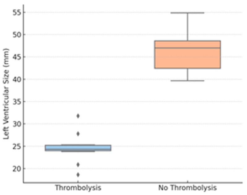
Objectives: Post-acute venous thromboembolism (VTE) is a well-recognized complication of COVID-19, driven by persistent endothelial dysfunction and thromboinflammation. Identifying simple clinical predictors of VTE may optimize therapy and limit adverse outcomes. We propose a pragmatic risk-stratification approach, based on clinical and echocardiographic parameters. Methods: We conducted a retrospective cohort study in a Romanian tertiary hospital (March 2020-April 2022) in 54 adults with laboratory-confirmed COVID-19 and imaging-confirmed VTE. Demographics, comorbidities, laboratory markers, and echocardiographic variables-particularly tricuspid annular plane systolic excursion (TAPSE), peripheral oxygen saturation (SpO2), and left-ventricular end-diastolic diameter (LVEDD)-were collected. The primary outcome was the percentage of patients receiving systemic thrombolysis. Statistical analyses included Mann-Whitney U tests, chi-square, Spearman correlations, and multivariable logistic regression. Results: The mean age was 61.2 ± 14.7 years, and 63% were men. Eleven patients (20.4%) underwent thrombolysis. Compared with conservatively managed patients, those receiving thrombolysis had lower TAPSE (13.0 vs. 20.8 mm), lower SpO2 (90.1 vs. 97.0%), and smaller LVEDD (24.4 vs. 46.1 mm); all differences were statistically significant. Each 1 mm decrease in TAPSE and 1% decrease in SpO2 increased the likelihood of thrombolysis (adjusted odds ratios 1.58 and 1.34, respectively). Inflammatory markers and right-ventricular diameter were not associated with treatment. Conclusions: Reduced TAPSE, lower SpO2, and decreased LVEDD identify post-COVID VTE patients at elevated risk of hemodynamic compromise requiring thrombolysis. A point-of-care assessment incorporating these variables may improve early risk stratification and guide therapeutic decisions.
Keywords: COVID-19; SpO2; TAPSE; VTE; echocardiography; post-COVID syndrome; pulmonary embolism; thrombolysis.
Conflict of interest statement
The authors declare no conflicts of interest.
Figures


References
-
- Raskob G.E., Angchaisuksiri P., Blanco A.N., Buller H., Gallus A., Hunt B.J., Hylek E.M., Kakkar A., Konstantinides S.V., McCumber M., et al. Thrombosis: A major contributor to global disease burden. Arterioscler. Thromb. Vasc. Biol. 2014;34:2363–2371. doi: 10.1161/ATVBAHA.114.304488. - DOI - PubMed
-
- Kearon C. Epidemiology of venous thromboembolism. Semin. Vasc Med. 2001;1:7–26. - PubMed
-
- Boyd S., Sheng Loh K., Lynch J., Alrashed D., Muzzammil S., Marsh H., Masoud M., Bin Ihsan S., Martin-Loeches I. The Incidence of Venous Thromboembolism in Critically Ill Patients with SARS-CoV-2 Infection Compared with Critically Ill Influenza and Community-Acquired Pneumonia Patients: A Retrospective Chart Review. Med. Sci. 2022;10:30. - PMC - PubMed
LinkOut - more resources
Full Text Sources
Miscellaneous
