. 2025 Aug 27;15(9):1243.
doi: 10.3390/biom15091243.

CD45 and Basigin (CD147) Are Functional Ligands for Galectin-8 on Human Leukocytes

Affiliations

CD45 and Basigin (CD147) Are Functional Ligands for Galectin-8 on Human Leukocytes

Jean-Philippe F Gourdine et al. Biomolecules. .

Abstract

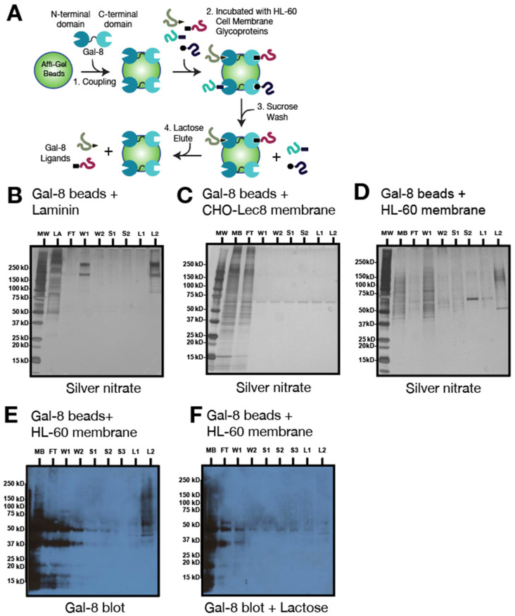
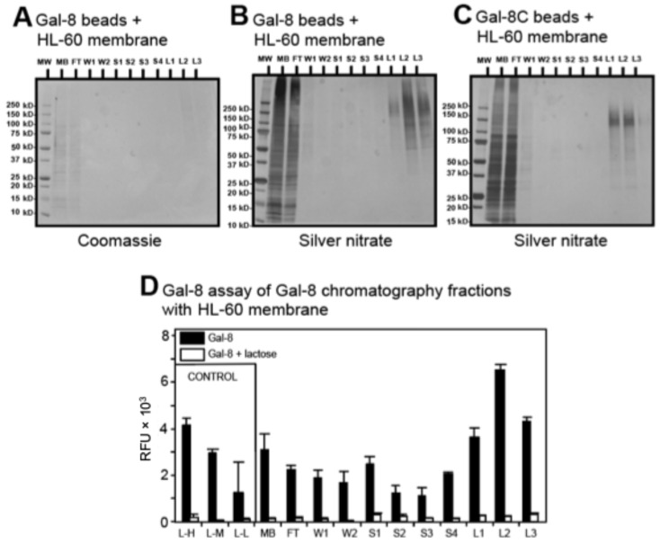
The interactions of leukocyte glycoproteins with adhesion and signaling molecules through glycan recognition are not well understood. We previously demonstrated that galectin-8, a tandem-repeat lectin with N- and C-terminal carbohydrate binding domains which is highly expressed in endothelial and epithelial cells, can bind to activated neutrophils to induce surface exposure of phosphatidylserine (PS) without DNA fragmentation or apoptosis, in a process termed preaparesis. However, the receptors for Gal-8 on leukocytes have not been identified. Here we report our results using both proteomics and affinity chromatography with both full-length Gal-8 and the separate Gal-8 C-terminal and N-terminal domains to identify glycoprotein ligands in HL-60 cells for Gal-8. Two of the major ligands for Gal-8 are CD45RA and CD45RC (Protein Tyrosine Phosphatase, PTP) and basigin (CD147). Both CD45 and basigin are integral membrane glycoproteins that carry poly-N-acetyllactosamine modifications on N- and/or O-glycans, required for Gal-8 binding. Inhibition of the phosphatase activity of CD45 reduced Gal-8-induced PS exposure, indicating a possible role of CD45 in Gal-8 signaling of preaparesis in human leukocytes. These results demonstrate unique glycoprotein recognition by Gal-8 involved in cell recognition and signaling.

Keywords: CD45; basigin; galectin-8; glycan ligands; leukocytes.


Conflict of interest statement

Author Alexander J. Noll was employed by The Emmes Company, LLC. The remaining authors declare that the research was conducted in the absence of any commercial or financial relationships that could be construed as a potential conflict of interest.

Figures

Figure 1
Figure 1
Validation of Gal-8 chromatography columns. (A) Schematic representation of Gal-8 chromatography with HL-60 cell membrane preparations and steps in the procedure. Gal-8 chromatography (1 mg Gal-8/mL beads) with (B), positive control: mouse laminin (LA); (C) negative control: CHO-Lec8 cell mutant membrane (250 μg/mL); (D) HL-60 cell membrane extract (250 μg/mL). (BD) were visualized with silver nitrate staining. (E,F) Blotting of fractions from (D) with biotinylated Gal-8 (1 μg/mL) in absence (E) and presence (F) of lactose. Legend: MW, molecular weight markers; LA, laminin; MB, total membrane fraction; FT, flow through; W, wash; S, 100 mM sucrose wash; L, 100 mM lactose elution.
Figure 2
Figure 2
Gal-8 chromatography with intact HL-60 cells. (A) Schematic representation of Gal-8 chromatography steps with intact HL-60 cells. (B) Gal-8 chromatography (1 mg Gal-8/mL beads) of intact, cell surface biotinylated HL-60 cells. Samples were visualized with silver nitrate staining. (C) Strep-HRP blot (1 μg/mL) of fractions from B with intact biotinylated HL-60 cells. Legend: MW, molecular weight markers; MB, total membrane fraction; FT, flow through; W, wash; S, 100 mM sucrose wash; L, 100 mM lactose elution.
Figure 3
Figure 3
Gal-8 binds to specific sets of glycoproteins. (A,B) High-density (10 mg Gal-8/mL beads) full-length Gal-8 chromatography with 8 mL of HL-60 cell membrane extract (250 μg/mL) visualized by Coomassie brilliant blue staining and silver nitrate, respectively; (C) high-density (10 mg Gal-8C/mL beads) Gal-8C domain chromatography with 8 mL of HL-60 cell membrane extract (250 μg/mL) visualized by silver nitrate; (D) Gal-8 binding to protein fractions eluted from Gal-8 chromatography fractions with HL-60 cell membrane preparation. Results represent mean of 2 independent experiments performed in triplicate. Error bars represent means ± 1 standard deviation (SD). Legend: MW, molecular weight markers; MB, total membrane fraction; FT, flow through; W, wash; S, 100 mM sucrose wash; L, 100 mM lactose elution; L-H, laminin at high amount (10 μg); L-M, laminin at medium amount (5 μg); L-L, laminin at low amount (1 μg).
Figure 4
Figure 4
CD45 was detected among the Gal-8 binding proteins on HL-60 cells. (A) Anti-CD45 blot of full-length Gal-8 chromatography with HL-60 cell membranes; (B) anti-CD45 blot of Gal-8C chromatography with HL-60 cell membranes; (C) Reverse Transcriptase-PCR of CD45 isoforms and beta-actin with HL-60 and Jurkat cells. Specific amplicons for CD45 (about 200, 350, and 500 bp) are visible; (D) HL-60 cell staining with anti-CD45-Alexa 488 or isotype control measured by flow cytometry; (E) confocal images of HL-60 cells treated at 4 °C with biotinylated Gal-8CM (top row) or Gal-8NM (bottom row) with Strep-Alexa 633 (red) and anti-CD45-Alexa 488 (green); Merge 1: whole cell reconstruction, Merge 2: detailed colocalization (yellow), scale bar = 5 μm. White arrows indicate colocalization in punctate membrane microdomains. Legend: MB, total membrane fraction; FT, flow through; W, wash; S, 100 mM sucrose wash; L, 100 mM lactose elution; MW, molecular weight.
Figure 5
Figure 5
Gal-8-induced phosphatidylserine (PS) exposure is blocked by inhibition of CD45 phosphatase activity. (A) Flow cytometry dot plot of HL-60 cells treated with 0.6 μM Gal-8NM final with or without 100 mM lactose (Lac) in presence of vehicle (DMSO) or CD45 inhibitor (CD45I, 1 μM); Cells were stained with Annexin-V (An-V) and propidium iodide (PI) to measure PS exposure (Annexin-V positive/PI negative); (B) PS exposure quantification of three independent experiments in duplicate of panel (A), p < 0.005 between DMSO-treated and CD45I-treated samples. Error bars represent means ± 1 SD of three independent experiments in duplicate; (C) Western blot detection of phospho-ERK and total ERK after 1 h incubation of HL-60 cells with Gal-8NM with or without lactose, and with DMSO or CD45 inhibitor (CD45I); (D) Western blot detection of p-ERK after 1 h incubation with Gal-8 with (+) or without (−) CD45I with increased loading of HL-60 cell lysate (2.5 to 20 μL).
Figure 6
Figure 6
Gal-8NM signaling is not inhibited by kifunensine (Kif) nor benzyl-αGalNAc (BG). (A,C) HL-60 cell staining with PHA-L (red), ConA (green), CHO-131-Alexa 488 (orange), Cholera toxin subunit B (blue), isotype control (grey), and Strep-Alexa 488 (black) measured by flow cytometry in presence or absence of inhibitors A: Kif or C: BG (solid lines) or absence (dotted-lines). (B,D) PS exposure quantification of three independent experiments in duplicate with Gal-8NM and Gal-1 in the presence of inhibitors (B): Kif or (D): BG (black) or absence (white). Error bars = means ± 1 SD.
Figure 7
Figure 7
Gal-8 binds to basigin through polyLacNAc present on N-glycans. (A) Gal-8 affinity chromatography of HL-60 cell membrane preparations. Blots were stained with anti-basigin (CD147) antibody, followed by HRP-conjugated goat anti-mouse, followed by ECL. (BD) Anti-basigin blot of full-length Gal-8 chromatography with HL-60 cell membranes; washes were performed with 3 mL of 100 mM sucrose (S) and elution with 3 mL of 100 mM lactose in PBS-TX; (A) mock-treated with buffers alone or (B), PNGase F-treated; (C) mock-treated with sodium acetate or (D), treated with endo-β-galactosidase (E. freundii). Legend: MB, total membrane fraction; FT, flow through; S, 100 mM sucrose wash; L, 100 mM lactose elution.
Figure 8
Figure 8
Both N- and C-terminal domains of Gal-8 bind to basigin and colocalize with basigin on HL-60 cells. (A) Anti-basigin (CD147) blot of Gal-8NM and Gal-8CM chromatography with HL-60 cell membranes (U: Unbound, B: Bound); (B) HL-60 cell staining with anti-basigin-Alexa 488 or isotype control measured by flow cytometry; (C) confocal images of HL-60 cells treated at 4 °C with (a) biotinylated Gal-8NM or (e) Gal-8CM with Strep-Alexa 633 (red) and (b,f) anti-basigin-Alexa 488 (green); Merged images Gal-8NM/anti-basigin and Gal-8CM/anti-basigin are shown (c,g), and zoomed images on two individual cells from (c,g) are shown (d,h), respectively. Scale bar represents 5 μm.
Figure 9
Figure 9
Working model of Gal-8-induced PS exposure on neutrophils. (1) Neutrophils can be activated after formyl-Methionyl-Leucyl-Phenylalanine (fMLP) receptor stimulation by degraded bacterial or mitochondrial proteins from dying surrounding cells; (2) Dimeric Gal-8 expressed by endothelial cells (ECs) or smooth muscle cells (SMCs) binds to polyLacNAc containing glycoproteins, particularly CD45, (3) which is upregulated during neutrophil activation [47]. CD45 signals through its phosphatase activity, dephosphorylating directly or indirectly phospho-proteins such as p-ERK, (4) leading to an increase of cytoplasmic calcium ions (Ca2+). This cation increase may be a synergistic result with fMLP receptor signaling through G-Proteins, which also conduct Protein kinase delta activation (PKCδ). PKCδ and Ca2+ will then (5) activate scramblase, which results in PS exposure [13,97]. Synergistically, dimeric Gal-8 could also bind to a CD147-Xkr8 complex, and possibly directly activate scramblase activity by Xkr8 [91]. ?? represents an unknown component of the process.

References

    1. Liu F.T., Stowell S.R. The role of galectins in immunity and infection. Nat. Rev. Immunol. 2023;23:479–494. doi: 10.1038/s41577-022-00829-7. - DOI - PMC - PubMed
    1. Zhang N., Liu Q., Wang D., Wang X., Pan Z., Han B., He G. Multifaceted roles of Galectins: From carbohydrate binding to targeted cancer therapy. Biomark. Res. 2025;13:49. doi: 10.1186/s40364-025-00759-1. - DOI - PMC - PubMed
    1. Cummings R.D., Rabinovich G.A., Stowell S.R., Vasta G. Galectins. In: Varki A., Cummings R.D., Esko J.D., Stanley P., Hart G.W., Aebi M., Mohnen D., Kinoshita T., Packer N.H., Prestegard J.H., et al., editors. Essentials of Glycobiology. 4th ed. Cold Spring Harbor Laboratory Press; Cold Spring Harbor, NY, USA: 2022.
    1. Stowell S.R., Arthur C.M., Dias-Baruffi M., Rodrigues L.C., Gourdine J.P., Heimburg-Molinaro J., Ju T., Molinaro R.J., Rivera-Marrero C., Xia B., et al. Innate immune lectins kill bacteria expressing blood group antigen. Nat. Med. 2010;16:295–301. doi: 10.1038/nm.2103. - DOI - PMC - PubMed
    1. Barondes S.H., Cooper D.N., Gitt M.A., Leffler H. Galectins. Structure and function of a large family of animal lectins. J. Biol. Chem. 1994;269:20807–20810. doi: 10.1016/S0021-9258(17)31891-4. - DOI - PubMed

LinkOut - more resources