. 2025 Sep 12;17(18):2988.
doi: 10.3390/cancers17182988.

Diagnostic Biomarker Candidates Proposed Using Targeted Lipid Metabolomics Analysis of the Plasma of Patients with PDAC

Affiliations

Diagnostic Biomarker Candidates Proposed Using Targeted Lipid Metabolomics Analysis of the Plasma of Patients with PDAC

Sung-Sik Han et al. Cancers (Basel). .

Abstract

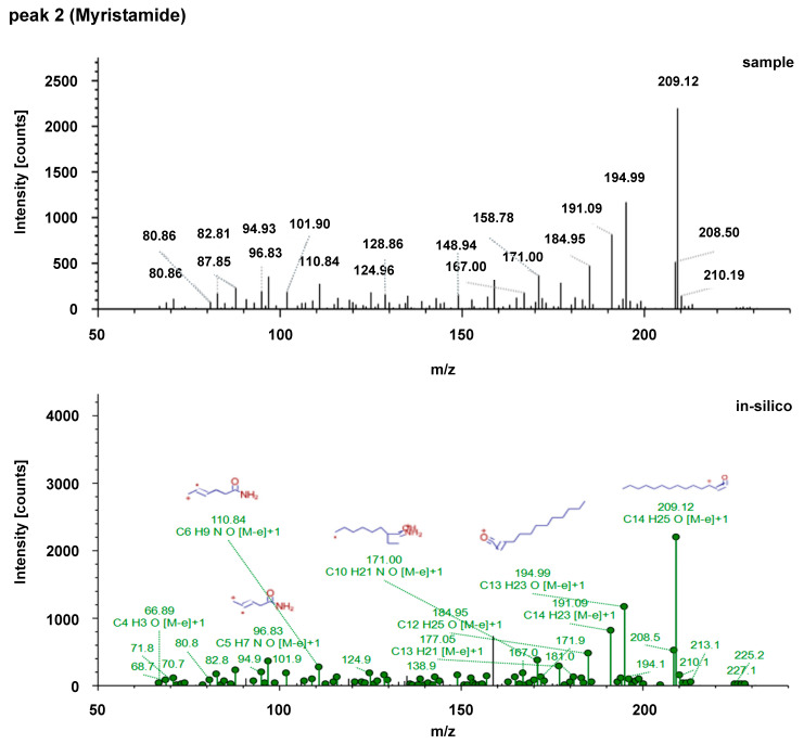
Background/Objectives: We recently discovered that tumors rely on blood fatty acids as an energy source for growth. Therefore, we investigated biomarkers in the lipid fractions of plasma from patients with pancreatic ductal adenocarcinoma (PDAC) for the screening diagnosis of PDAC. Methods: We screened common fatty acid types in human (normal 99, PDAC 103) and mouse (normal 7, KPC 22) plasma samples using a non-targeted approach. Subsequently, we identified targets in human plasma (set A: normal 68, PC 102) that could distinguish between healthy individuals and patients with cancer. Next, we verified whether the identified targets were useful in a new human set (set B: 96 normal, 78 PC). We combined sets A and B to create set C and further divided it into a training set (7:3 ratio; normal 115, pancreatic cancer 126) and a validation set (normal 49, PC 54). The identified targets were used to train three statistical models (logistic regression (LR), random forest (RF), and support vector machine (SVM) with a radial basis function (RBF) kernel). Results: The comparison of human and mouse plasma identified eight common lipid metabolites. We further identified four platforms containing these metabolites for target analysis: acylcarnitines, phospholipids, fatty acid amides, and sphingolipids. We analyzed the four platforms using sets A, B, and C and found 20 lipids (1 acylcarnitine, 1 sphingolipid, and 18 phospholipids) that met the criterion of AUC ≥ 0.75 in all three sets. Based on an average AUC for LR models with 11 or more phospholipids, the separation performance between healthy individuals and patients with cancer was 0.9207 (sensitivity, 90.74%; specificity, 86.22%; PPV, 87.90%; NPV, 89.42%), and the AUC of the validation set for CA19-9 in the same groups was 0.7354. The addition of CA19-9 to the LR models resulted in a separation performance of 0.9427 (90.74%; 88.01%; 89.32%; 89.61%) for the validation set. Conclusions: We identified 18 candidate fatty acid metabolites that could serve as biological markers in the serum lipid fractions of pancreatic cancer patients and confirmed that all of them decreased in patients. Additionally, we developed an algorithm utilizing these markers, which demonstrated a 25% increase in discriminatory power compared to the AUC value of CA19-9, an FDA-approved biomarker for pancreatic cancer. In summary, we identified candidate metabolites and algorithms that could serve as biomarkers in the lipid fractions of plasma from patients with pancreatic cancer.

Keywords: CA19-9; diagnostic biomarker; lipid metabolomics; pancreatic cancer.


Conflict of interest statement

Authors Jun Hwa Lee, Joon Hee Kang, and Soo-Youl Kim hold stocks of NCC-Bio Co. Author Sang Myung Woo has been involved as a consultant and expert witness in NCC-Bio Co. Authors Sung-Sik Han, Sang Myung Woo, Jun Hwa Lee, Sang-Jae Park, Woo Jin Lee, Kyung-Hee Kim, and Soo-Youl Kim are the inventors of the patent (Composition of biomarkers for cancer diagnosis including geochemical metabolites and uses thereof, KR 10-2023-0053911).

Figures

Figure A1
Figure A1
MS/MS spectra of the eight common metabolites in the positive mode in Table 2.
Figure A1
Figure A1
MS/MS spectra of the eight common metabolites in the positive mode in Table 2.
Figure A1
Figure A1
MS/MS spectra of the eight common metabolites in the positive mode in Table 2.
Figure A1
Figure A1
MS/MS spectra of the eight common metabolites in the positive mode in Table 2.
Figure A1
Figure A1
MS/MS spectra of the eight common metabolites in the positive mode in Table 2.
Figure A1
Figure A1
MS/MS spectra of the eight common metabolites in the positive mode in Table 2.
Figure A1
Figure A1
MS/MS spectra of the eight common metabolites in the positive mode in Table 2.
Figure A1
Figure A1
MS/MS spectra of the eight common metabolites in the positive mode in Table 2.
Figure A2
Figure A2
AUC plots of random forest (RF) and support vector machine (SVM) models with cumulative addition of phospholipids arranged in descending order of the AUC: (A) RF without CA19-9, (B) RF with CA19-9, (C) SVM without CA19-9, and (D) SVM with CA19-9.
Figure 1
Figure 1
Strategy for deriving candidate lipid metabolites for screening patients with PDAC. Derivation of common target lipid metabolites through non-targeted metabolomics analysis of the plasma of human patients with cancer and a mouse cancer model using human PDAC cells.
Figure 2
Figure 2
Extracted ion chromatograms and peak areas of human and mouse plasma samples. (A) The ion chromatogram revealed distinct metabolomic features in PC human and KPC mouse plasma compared to their matched controls. (B) Peak areas of human and mouse plasma samples, along with corresponding ROC curves of the 5th common metabolomic feature: Upper left panel: peak areas of human plasma samples, red dot-pancreatic cancer patients, blue dot-normal persons. Upper right panel: ROC curve of human plasma samples, Lower left panel: peak areas of mouse plasma samples, red dot-KPC mice, blue dot-normal mice, and Lower right panel: ROC curve of mouse plasma samples.
Figure 2
Figure 2
Extracted ion chromatograms and peak areas of human and mouse plasma samples. (A) The ion chromatogram revealed distinct metabolomic features in PC human and KPC mouse plasma compared to their matched controls. (B) Peak areas of human and mouse plasma samples, along with corresponding ROC curves of the 5th common metabolomic feature: Upper left panel: peak areas of human plasma samples, red dot-pancreatic cancer patients, blue dot-normal persons. Upper right panel: ROC curve of human plasma samples, Lower left panel: peak areas of mouse plasma samples, red dot-KPC mice, blue dot-normal mice, and Lower right panel: ROC curve of mouse plasma samples.
Figure 3
Figure 3
Venn diagram of lipids with AUC ≥ 0.75 in sets A, B, and C.
Figure 4
Figure 4
AUC plots of logistic regression models with the cumulative addition of phospholipids arranged in descending order of the AUC (A) without CA19-9 and (B) with CA19-9.

References

    1. Bengtsson A., Andersson R., Ansari D. The actual 5-year survivors of pancreatic ductal adenocarcinoma based on real-world data. Sci. Rep. 2020;10:16425. doi: 10.1038/s41598-020-73525-y. - DOI - PMC - PubMed
    1. Siegel R.L., Miller K.D., Wagle N.S., Jemal A. Cancer statistics, 2023. CA Cancer J. Clin. 2023;73:17–48. doi: 10.3322/caac.21763. - DOI - PubMed
    1. Yin X., Xu R., Song J., Ruze R., Chen Y., Wang C., Xu Q. Lipid metabolism in pancreatic cancer: Emerging roles and potential targets. Cancer Commun. 2022;42:1234–1256. doi: 10.1002/cac2.12360. - DOI - PMC - PubMed
    1. Su S.B., Qin S.Y., Chen W., Luo W., Jiang H.X. Carbohydrate antigen 19-9 for differential diagnosis of pancreatic carcinoma and chronic pancreatitis. World J. Gastroenterol. 2015;21:4323–4333. doi: 10.3748/wjg.v21.i14.4323. - DOI - PMC - PubMed
    1. Fahrmann J.F., Schmidt C.M., Mao X., Irajizad E., Loftus M., Zhang J., Patel N., Vykoukal J., Dennison J.B., Long J.P., et al. Lead-Time Trajectory of CA19-9 as an Anchor Marker for Pancreatic Cancer Early Detection. Gastroenterology. 2021;160:1373–1383.e6. doi: 10.1053/j.gastro.2020.11.052. - DOI - PMC - PubMed

LinkOut - more resources