

Contrast-Enhanced Mammography-Guided Biopsy in Patients with Extensive Suspicious Microcalcifications
- PMID: 41008928
- PMCID: PMC12468424
- DOI: 10.3390/cancers17183086
Contrast-Enhanced Mammography-Guided Biopsy in Patients with Extensive Suspicious Microcalcifications
Abstract
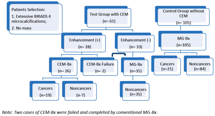
Objectives: To investigate the feasibility of contrast-enhanced mammography-guided biopsy (CEM-Bx) to diagnose cancer via targeting the associated enhancements in the patients with extensive suspicious microcalcifications. Methods: All the women with extensive suspicious microcalcifications were mammographically screened. Contrast-enhanced mammography was first examined, followed by CEM-Bx if there was any relevant enhancement; otherwise, patients without enhancement were submitted to conventional mammography-guided biopsy (MG-Bx). We recorded and analyzed the histological results, morphologies and distributions of the microcalcifications. The outcomes were also compared to those patients (control group) who did not assess with CEM and received MG-Bx only by the Wilcoxon rank-sum test. Results: Between November 2021 and November 2023, a total of 61 participants participated in the test. A total of 26 women underwent CEM-Bx, and 35 underwent MG-Bx. In total, 19 of the 26 CEM-Bx were diagnosed as cancer, and none by MG-Bx. The cancer diagnostic rates (CDRs) identified by CEM-Bx were 81.8% for regional microcalcifications and 66.7% for segmental or diffuse distributions. The CDR of the test group was higher than the control group, 31.4% to 20%, respectively. Otherwise, the CDR of CEM-Bx was significantly higher than MG-Bx in the control group (73.08% to 20%, p-valve < 0.01). Conclusions: CEM-Bx was a safe and feasible procedure. With identification of the enhanced target, CEM-Bx faithfully performed among the extensive distributed suspicious microcalcifications. Although CEM-Bx improves CDR, larger prospective trials with surgical validation of all lesions are needed before widespread adoption.
Keywords: breast cancer; breast microcalcifications; contrast-enhanced mammography; contrast-enhanced mammography guided biopsy; diagnosis; mammography.
Conflict of interest statement
Author Chia-Wei Li was employed by the company GE HealthCare. The remaining authors declare that the research was conducted in the absence of any commercial or financial relationships that could be construed as a potential conflict of interest.
Figures

References
-
- Catanzariti F., Avendano D., Cicero G., Garza-Montemayor M., Sofia C., Rullo E.V., Ascenti G., Pinker-Domenig K., Marino M.A. High-risk lesions of the breast: Concurrent diagnostic tools and management recommendations. Insights Imaging. 2021;12:63. doi: 10.1186/s13244-021-01005-6. - DOI - PMC - PubMed
Grants and funding
LinkOut - more resources
Full Text Sources
