Review
. 2025 Aug 23;14(9):1041.
doi: 10.3390/antiox14091041.

Antioxidant Molecules in the Human Vitreous Body During Prenatal Eye Development

Affiliations
Review

Antioxidant Molecules in the Human Vitreous Body During Prenatal Eye Development

Ina G Panova et al. Antioxidants (Basel). .

Abstract

The structures of the developing eye may be damaged as a result of the impact of reactive oxygen species (ROS) interacting with different cellular components. The antioxidant molecules found in the eye, especially in the vitreous body-the largest component of the eye, playing a crucial role in the formation of structures and functions of the developing eye-provide protection to the eye tissues from ROS. This review considers various antioxidant molecules (ascorbic acid, lutein, bilirubin, uric acid, catecholamines, erythropoietin, albumin, and alpha-fetoprotein) that have been found in the human vitreous body during the early stages of pregnancy (10-31 weeks of gestation) and their functions in the development of the eye. The presence of some molecules is transient (lutein, AFP), whereas a temporal decrease (albumin, bilirubin) or increase (ascorbic acid, erythropoietin) in the concentrations of other antioxidants is observed. Since the actual overall content of antioxidants in the developing vitreous body is probably much higher than that found to date, further research is needed to study antioxidants there. It is especially important to study the antioxidant status of the vitreous body at the earliest stages of its development. Antioxidants found suggest their use for the prophylactic of ocular diseases during pregnancy and finding new antioxidants could create an additional opportunity in this regard.

Keywords: antioxidants; human eye; prenatal development; reactive oxygen species; vitreous body.


Conflict of interest statement

The authors declare that they have no competing interests.

Figures
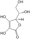
Figure 1
Figure 1
Structure of vitamin C (L-ascorbic acid), an active antioxidant. Due to the presence of four OH groups, it is soluble in water.
Figure 2
Figure 2
Structure of lutein. Due to the presence of a long polyenic chain, lutein effectively quenches singlet oxygen and is an active antioxidant.
Figure 3
Figure 3
Redox equilibrium bilirubin–biliverdin, which determines bilirubin antioxidant activity. Two meso-H atoms of a bilirubin molecule easily react with ROS, converting them into inactive forms.
Figure 4
Figure 4
Structure of uric acid. As an antioxidant, it enters into one-electron redox reaction with active oxy radicals and effectively quenches singlet oxygen.
Figure 5
Figure 5
Structures of epinephrine (adrenaline) (a), norepinephrine (noradrenaline) (b), and dopamine (c). Their antioxidant properties are mostly determined by a catechol (phenol) group.
Figure 6
Figure 6
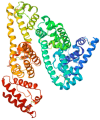
Structure of human EPO molecule (from [146]), an active antioxidant. It contains three N-linked and one O-linked side chains of acidic oligosaccharides.
Figure 7
Figure 7
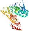
Structure of HSA—the main protein in the body (from [177]). It mainly performs transport functions and is also an antioxidant.
Figure 8
Figure 8
Structure of AFP molecule (from [197]). As HSA, it carries out transport and antioxidant functions.
Figure 9
Figure 9
Stages of development of the vitreous (according to Ida Mann [11]) and its molecular components with their timing. Collagens [21], chondroitin-sulfate-proteoglycans [21], hyaluronic acid [21,23], and opticin [24] are structural components of the vitreous and are not antioxidants. Albumin [180,181,182], alpha-fetoprotein [182,198], erythropoietin [172], ascorbic acid [58], lutein [76], bilirubin [117], uric acid [128], and catecholamines [128] have antioxidant properties. N/s, not studied; N/f, not found.
Figure 10
Figure 10
Synergistic relationship between the antioxidants found in the fetal vitreous. Ascorbic acid reduces the oxidized urate radical formed in reactions with ROS, which repairs the initial uric acid molecule (label “R”). Ascorbic acid also stimulates the synthesis of neuronal catecholamines, which increases their antioxidative effect (label “S”). Albumin and alpha-fetoprotein have transport functions with respect to several antioxidants, in particular, bilirubin and lutein, delivering them to target tissues (label “Transport”).

References

    1. Balazs E.A., Toth L.Z., Ozanics V. Cytological studies on the developing vitreous as related to the hyaloid vessel system. Graefe’s Arch. Clin. Exp. Opthalmol. 1980;213:71–85. doi: 10.1007/BF00413534. - DOI - PubMed
    1. Bishop P.N. Structural macromolecules and supramolecular organization of the vitreous gel. Prog. Retin. Eye. Res. 2000;19:323–344. doi: 10.1016/S1350-9462(99)00016-6. - DOI - PubMed
    1. Bishop P.N., Takanosu M., le Goff M., Mayne R. The role of the posterior ciliary body in the biosynthesis of vitreous humour. Eye. 2002;16:454–460. doi: 10.1038/sj.eye.6700199. - DOI - PubMed
    1. Hogan V.J., Alvarado J.A., Weddel J.E. Histology of the Human Eye. W.B. Saunders Company; Philadelphia, PA, USA: 1971. 687р
    1. Ankamah E., Sebag J., Ng E., Nolan J.M. Vitreous antioxidants, degeneration, and vitreo-retinopathy: Exploring the links. Antioxidants. 2020;9:7. doi: 10.3390/antiox9010007. - DOI - PMC - PubMed

LinkOut - more resources