. 2025 Sep 5;16(9):1045.
doi: 10.3390/genes16091045.

Teashirt and C-Terminal Binding Protein Interact to Regulate Drosophila Eye Development

Affiliations

Teashirt and C-Terminal Binding Protein Interact to Regulate Drosophila Eye Development

Surya Jyoti Banerjee et al. Genes (Basel). .

Abstract

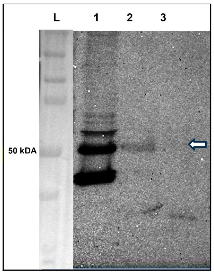
Background and Objectives: The Drosophila retinal determination network comprises the transcription factor Teashirt (Tsh) and the transcription co-regulator C-terminal Binding Protein (CtBP), both of which are essential for normal adult eye development. Both Tsh and CtBP show a pattern of co-expression in the proliferating cells anterior to the morphogenetic furrow that demarcates the boundary between the anteriorly placed proliferating eye precursor cells and the posteriorly placed differentiating photoreceptor cells in the larval eye-precursor tissue, the eye-antennal disc. The disc ultimately develops into the adult compound eyes, antenna, and other head structures. Both Tsh and CtBP were found to interact genetically during ectopic eye formation in Drosophila, and both were present in molecular complexes purified from gut and cultured cells. However, it remained unknown whether Tsh and CtBP molecules could interact in the eye-antennal discs and elicit an effect on eye development. The present study answers these questions. Methods: 5' GFP-tagging of the tsh gene in the Drosophila genome and 5' FLAG-tagging of the ctbp gene were accomplished by the CRISPR-Cas9 and BAC recombineering methods, respectively, to produce GFP-Tsh- and FLAG-CtBP-fused proteins in specific transgenic Drosophila strains. Verification of these proteins' expression in the larval eye-antennal discs was performed by immunohistological staining and confocal microscopy. Genetic screening was performed to establish functional interaction between Tsh and CtBP during eye development. Scanning Electron Microscopy was performed to image the adult eye structure. Co-immunoprecipitation and GST pulldown assays were performed to show that Tsh and CtBP interact in the cells of the third instar eye-antennal discs. Results: This study reveals that Tsh and CtBP interact genetically and physically in the Drosophila third instar larval eye-antennal disc to regulate adult eye development. This interaction is likely to limit the population of the eye precursor cells in the larval eye disc of Drosophila. Conclusions: The relative abundance of Tsh and CtBP in the third instar larval eye-antennal disc can dictate the outcome of their interaction on the Drosophila eye formation.

Keywords: BAC recombineering; C-terminal binding protein; CRISPR-Cas9; Drosophila; GST pulldown; co-immunoprecipitation; eye development; genetic interaction; teashirt.


Conflict of interest statement

The authors declare no conflicts of interest.

Figures

Figure 1
Figure 1
Confocal images of the third instar larval eye–antennal discs showing Tsh and Elav expression patterns (the anterior side is on the left). The top and bottom panels contain the confocal images of the third instar larval eye–antennal discs dissected from the EGFP-tsh (A1A3) and the BL# 52669 (B1B3) (negative control) flies, respectively. The eye–antennal disc from the EGFP-tsh fly showed Tsh expression (identified by GFP expression) in the anterior part (to the left) of the eye disc (A1), which was absent in the control disc (B1). Elav expression was seen at the posterior part of both the eye discs (A2,B2). In the merged images (A3,B3), the morphogenetic furrow could be seen as an unstained region between the cells expressing Tsh (green) and Elav (red) in (A3).
Figure 2
Figure 2
Confocal images of the third instar larval eye–antennal discs showing CtBP, Elav, and Ey expression patterns (the anterior side is on the left). The top and bottom panels contain the confocal images of the third instar larval eye–antennal discs dissected from the FLAG-ctbp (shown as CtBP-FLAG) (A1A3) and Ey-FLAG (positive control) (B1B3) flies, respectively. The eye–antennal disc from the FLAG-ctbp fly showed CtBP expression (identified by FLAG expression) in the anterior part of the eye disc and the entire antennal disc (A1). The eye–antennal disc from the Ey-FLAG fly showed Ey expression (identified by FLAG expression) in the anterior part of the eye disc (B1). Elav expression was seen at the posterior part of both the eye discs (A3,B3). In the merged images (A2,B2), the morphogenetic furrow could be seen as an unstained region between the cells expressing CtBP (pink) and Elav (green) in A2; and Ey (green) and Elav (pink) (B2).
Figure 3
Figure 3
Light microscopy images of adult eyes of flies having up- and downregulation of tsh and ctbp, and respective controls. The adult eyes of the control flies, including the eyeless-Gal4 (A,F) driver, and UAS-tsh (B); UAS-tshIR (D); UAS-ctbp ORF (G); and UAS-ctbpIR (I) were compared with the adult eyes of their progenies (i) overexpressing tsh (ey > tsh) (C) and ctbp (ey > ctbp ORF) (H); and progenies having downregulation of (ii) tsh (ey > tshIR) (E) or ctbp (ey > ctbpIR) (J) expression. The (C) ey > tsh flies lack the entire eyes or had tiny eyes; the (E) ey > tshIR flies had no eye phenotype; the (H) ey > ctbp ORF flies had smaller eyes; and the (J) ey > ctbpIR flies had slightly bigger eyes relative to their parents’ eyes.
Figure 4
Figure 4
Scanning electron micrographs of adult fly eyes show that the genetic interaction of tsh with ctbp in the eye–antennal disc regulates eye development. Overexpression of tsh (A2,A3) but not GFP (A1) by the ey-Gal4 driver produced no or tiny adult eyes. These no or tiny eye phenotypes are partially rescued in flies also having a LoF tsh allele (tsh8) (B2) or ctbp downregulation (B3). The control flies containing a LoF tsh allele (tsh8/CyO) (B1) or UAS-ctbpIR (Figure 3I) had normal adult eyes.
Figure 5
Figure 5
Scanning electron micrograph showing that the small or misshapen adult eye phenotypes generated by overexpression of ctbp were rescued by removing a functional allele of tsh. Overexpression of ctbp ORF (A2) but not GFP (A1) by the ey-Gal4 driver produced small adult eyes (A2). The small eye phenotype was rescued in the flies also having a LoF tsh allele (tsh8) (A3). Overexpression of ctbp (B2) (a different UAS-ctbp line with the P[GSV]A396) but not GFP (B1) by the ey-Gal4 driver produced adult eyes with lost ommatidia at the edges (A2). The absence of ommatidia at the edges was rescued in the flies also having a LoF tsh allele (tsh8) (B3). The control flies containing a LoF tsh allele (tsh8) (B1) or P[GSV]A396 [22] had normal eyes.
Figure 6
Figure 6
Co-immunoprecipitation showing Tsh pulls down CtBP from the lysate prepared from the third instar larval eye–antennal discs of EGFP-tsh transgenic flies. IP with anti-EGFP antibody and Western blot with anti-CtBP antibody using lysate prepared from third instar larval eye–antennal discs dissected from EGFP-tsh transgenic flies. The lane marked by “L” contains the protein ladders (BioRad Precision Plus Protein Dual Color Standards). A 50 kilo-Dalton (kDA) band representing CtBP was present in the crude lysate (lane 1). A similar-sized band was precipitated by anti-EGFP antibody (lane 2) but not by the Protein G beads (negative control) (lane 3). The white arrow marks the 50kDA band size.
Figure 7
Figure 7
GST pulldown experiments reveal that Tsh and CtBP proteins bind directly. (A) shows that the GST-CtBP, but not the GST protein, can pull down the Tsh protein. The first lane contains the 10% sample from the in vitro translated Tsh prey protein band marked as “Load”, and the protein band in the third lane represents Tsh, pulled down by the GST-CtBP bait protein. The absence of the same band in the center lane signifies that GST alone could not pull down Tsh, suggesting CtBP binds and pulls down Tsh. (B) shows that the GST-Tsh, but not the GST protein, can pull down the CtBP protein. The first lane contains the 10% sample from the in vitro translated CtBP prey protein band marked as “Load”, and the protein band in the third lane represents CtBP, pulled down by the GST-Tsh bait protein. The absence of the same band in the center lane signifies that GST alone could not pull down CtBP, suggesting Tsh binds and pulls down CtBP.

References

    1. Silver S.J., Rebay I. Signaling circuitries in development: Insights from the retinal determination gene network. Development. 2005;132:3–13. doi: 10.1242/dev.01539. - DOI - PubMed
    1. Baker N.E., Firth L.C. Retinal determination genes function along with cell-cell signals to regulate Drosophila eye development: Examples of multi-layered regulation by master regulators. Bioessays. 2011;33:538–546. doi: 10.1002/bies.201000131. - DOI - PMC - PubMed
    1. Pappu K., Mardon G. Results and Problems in Cell Differentiation. Volume 37. Springer; Berlin/Heidelberg, Germany: 2002. Retinal specification and determination in Drosophila; pp. 5–20. - PubMed
    1. Fristrom D., Fristrom J.W. The Metamorphic Development of the Adult Epidermis. Volume 1–2 CSH Press; New York, NY, USA: 1993.
    1. Kumar J.P. Retinal determination the beginning of eye development. Curr. Top. Dev. Biol. 2010;93:1–28. - PMC - PubMed

MeSH terms

LinkOut - more resources