. 2025 Sep 11;13(9):2130.
doi: 10.3390/microorganisms13092130.

Biosurfactant-Mediated Inhibition of Salmonella Typhimurium Biofilms on Plastics: Influence of Lipopolysaccharide Structure

Affiliations

Biosurfactant-Mediated Inhibition of Salmonella Typhimurium Biofilms on Plastics: Influence of Lipopolysaccharide Structure

Shadi Khonsari et al. Microorganisms. .

Abstract

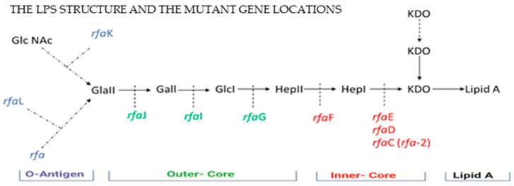
Salmonella enterica subsp. enterica serovar Typhimurium is a major foodborne pathogen whose ability to form biofilms contributes to persistent contamination in food-processing and clinical environments. This study investigated the anti-biofilm activity of the biosurfactant surfactin, produced by Bacillus subtilis, against S. Typhimurium wild type (LT2) and its lipopolysaccharide (LPS)-modified mutants on commonly used plastic surfaces such as polypropylene (PP) and polystyrene (PS). Biofilm formation was quantified using the crystal violet assay, revealing significantly higher biomass on PS compared to PP (p < 0.0001). Surfactin at 5 µg/mL was identified as the minimum biofilm inhibitory concentration (MBIC), significantly reducing biofilm formation in the wild-type and LPS mutants rfaL, rfaJ, rfaF (all p < 0.0001), and rfaI (p < 0.01). Further analysis using fluorescence microscopy and SYPRO® Ruby staining confirmed a significant reduction in extracellular polymeric substances (EPSs) on PP surfaces following surfactin treatment, particularly in strains LT2 (p < 0.0001), rfa (p < 0.01), rfaL (p < 0.0001), rfaG (p < 0.05), and rfaE (p < 0.0001). These findings highlight the influence of LPS structure on biofilm development and demonstrate surfactin's potential as an eco-friendly antimicrobial agent for controlling S. Typhimurium biofilms on food-contact surfaces. Analysis of mutants revealed that disruption of the rfa gene, which is involved in the biosynthesis of the outermost region of the lipopolysaccharide (LPS), significantly reduced bacterial attachment to polypropylene. This suggests that interactions between the external LPS layer and the plastic surface are important for colonisation. In contrast, mutants in core LPS biosynthesis genes such as rfaE and rfaD did not show any notable differences in attachment compared to the wild-type strain. This highlights the specific importance of outer LPS components, particularly under surfactant conditions, in mediating interactions with plastic surfaces. This work supports the application of biosurfactants in food safety strategies to reduce the risk of biofilm-associated contamination.

Keywords: Salmonella; biofilm; lipopolysaccharide; plastic surfaces; surfactin.


Conflict of interest statement

The authors declare no conflicts of interest.

Figures

Figure 1
Figure 1
Lipopolysaccharide structure of Gram-negative bacteria, including three regions located on the outer-membrane (OM) LPS. The main component of the OM can change its structure in response to the environment and strongly stimulate the innate immune response, which is also crucial in host–pathogen interactions [12].
Figure 2
Figure 2
Structure and biosynthesis pathway of lipopolysaccharide (LPS) in S. Typhimurium, illustrating the major LPS components (in black) and the specific biosynthesis genes (in different colours) involved at each stage of LPS assembly (Modified from [25]). The biosynthesis involves sequential addition of sugars such as GlcNAc (N-acetylglucosamine), Gal (galactose), Glc (glucose), Hep (heptose), and KDO (3-deoxy-D-manno-oct-2-ulosonic acid) to a Lipid A anchor. The LPS-modified mutants used in this study are indicated in different colours based on their LPS locations: rfa, rfaL, rfaK, rfaJ, rfaI, rfaG, rfaF, rfaE, rfaD.
Figure 3
Figure 3
Biofilm formation by S. Typhimurium WT (LT2) and ten LPS-modified gene mutants with deletion of genes located on different parts of LPS. Mutants are presented in order of their location on LPS: O-antigen genes rfa, rfaL, rfaK; inner-core genes rfaJ, rfaGrfaI,; outer-core genes rfaF, rfaE, rfaD. ****: p < 0.0001.
Figure 4
Figure 4
(a) Inhibition of S. Typhimurium LT2 biofilm formations on PS surface by surfactin at a range of concentrations, from the lowest concentration of 0.005 µg/mL to the highest concentration of 100 µg/mL, data presented as % biofilm formation relative to untreated control. (b) S. Typhimurium LT2 was grown on XLD plates in the presence of 5 µg/mL surfactin. NTC indicates the LT2 has no added biosurfactant (negative control); T0 is the initial time when biosurfactant has been applied to the bacterial cells grown on XLD, and T1 is 4 h after the application of biosurfactant. **: p < 0.01, *** p < 0.001 ****: p < 0.0001, ns: no significant difference.
Figure 5
Figure 5
Biofilm formation inhibition in S. Typhimurium WT and mutants on polypropylene (PP) (a) and polystyrene (PS) (b) surfaces in the presence of 5 µg/mL. *: p < 0.05, **: p < 0.01, ****: p < 0.0001.
Figure 6
Figure 6
The EPS biofilms of strains formed on PP coupon surfaces, untreated and treated with the antimicrobial agents bleach and surfactin (from left to right), stained with FilmTracerTM SYPROTM Ruby Biofilm Matrix Stain observed under Olympus Bx51 Fluorescence Microscope; images obtained using AmScope MU900-CK Microscope Digital Camera (20×). Blank stained coupon used as the control to observe the coupon surface without biofilm cell. Representative images of n = 15. Fluorescence intensity (%) of EPS measured with Fiji. One-way ANOVA reveals significant reduction of EPS treated with bleach and surfactin; * p < 0.05, *** p < 0.001, **** p < 0.0001, ns: no significant difference. Graphs/Images: (a) S. Typhimurium WT, (b) mutant rfaL, (c) mutant rfaG, (d) mutant rfaE. Each bar from left to right: non-treated (N), bleach-treated (B), and surfactin-treated (S).

References

    1. Flemming H., Wuertz S. Bacteria and archaea on Earth and their abundance in biofilms. Nat. Rev. Microbiol. 2019;17:247–260. doi: 10.1038/s41579-019-0158-9. - DOI - PubMed
    1. Valvano M.A., Furlong S.E., Patel K.B. Genetics biosynthesis assembly of O-antigen. In: Knirel Y.A., Valvano M.A., editors. Bacterial Lipopolysaccharides: Structure, Chemical Synthesis, Biogenesis and Interaction with Host Cells. Springer; Vienna, Austria: 2011. pp. 275–310. - DOI
    1. Muhammad M.H., Idris A.L., Fan X., Guo Y., Yu Y., Jin X., Qiu J., Guan X., Huang T. Beyond Risk: Bacterial Biofilms and Their Regulating Approaches. Front. Microbiol. 2020;11:928. doi: 10.3389/fmicb.2020.00928. - DOI - PMC - PubMed
    1. Moser C., Pedersen H.T., Lerche C.J., Kolpen M., Line L., Thomsen K., Høiby N., Jensen P.Ø. Biofilms and host response–helpful or harmful. Apmis. 2017;125:320–338. doi: 10.1111/apm.12674. - DOI - PubMed
    1. Maldonado R.F., Sá-Correia I., Valvano M.A. Lipopolysaccharide modification in gram-negative bacteria during chronic infection. FEMS Microbiol. Rev. 2016;40:480–493. doi: 10.1093/femsre/fuw007. - DOI - PMC - PubMed

LinkOut - more resources