The carbonic anhydrase 3 protein in urine: a potential biomarker to monitor atherosclerotic renal artery stenosis
- PMID: 41013375
- PMCID: PMC12465833
- DOI: 10.1186/s12882-025-04457-w
The carbonic anhydrase 3 protein in urine: a potential biomarker to monitor atherosclerotic renal artery stenosis
Abstract
Background: Atherosclerotic renal artery stenosis (ARAS) represents the predominant etiology of renal artery stenosis. Current diagnostic modalities—including renal arteriography, Doppler ultrasonography (DUS), computed tomography angiography (CTA), and magnetic resonance angiography (MRA)—are limited by invasiveness or technical constraints. The development of noninvasive biomarkers for ARAS detection is therefore clinically imperative. We hypothesize that ischemic renal injury in ARAS induces pathological alterations that generate unique urinary protein signatures.
Methods: A total of 138 subjects were enrolled and randomly divided into the discovery cohort and the validation cohort. Urinary proteins from the discovery cohort were profiled using data-independent acquisition mass spectrometry (DIA-MS) to identify differentially expressed proteins. Candidate biomarkers were subsequently validated in the validation cohort via quantitative ELISA. Diagnostic performance was assessed by receiver operating characteristic (ROC) curve analysis, and associations between target protein levels and clinical parameters were evaluated using Spearman correlation.
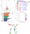
Results: As a result of DIA-MS indicated in the discovery cohort, 485 up-regulated urinary proteins and 177 down-regulated urinary proteins were identified in ARAS patients compared to disease controls. The top three proteins CA3, ORM2, and ART3 in AUC were selected as the potential urinary markers for further validation in the validation cohort. The uCA3/Cr level was significantly higher in both ARAS and disease controls than in healthy controls, and the uCA3/Cr level in patients with ARAS was significantly higher than in disease controls. There was no statistical difference in the uCA3/Cr level in subgroups with different severity of ARAS. Taking healthy controls as control values, the ROC area of uCA3/Cr was 0.9649(95%CI: 0.9200-1.000). We evaluated the area under the uCA3/Cr curve with the values of the disease control group as the control, the areas of ROC were 0.7391(95%CI: 0.6154–0.8628). Correlation analysis demonstrated that there was a strong positive correlation between uCA3/Cr concentration and urinary a1-microglobulin(a1MG) in ARAS and disease controls.
Conclusion: CA3 in urine may be related to renal interstitial injury, it could be used as a non-invasive biomarker for ARAS. However, large cohort studies and mechanistic investigations are required for further validation.
Supplementary Information: The online version contains supplementary material available at 10.1186/s12882-025-04457-w.
Keywords: Atherosclerosis renal artery stenosis; Biomarker; Carbonic anhydrase 3; Diagnosis; Urine proteomics.
Conflict of interest statement
Declarations. Ethics approval and consent to participate: The study was conducted in compliance with the declaration of Helsinki principles and followed the recommendations of Medical Ethics Committee of Aerospace Center. Hospital (20201113-QTKT-01) and all subjects provided written consent before inclusion. Consent for publication: Not applicable. Competing interests: The authors declare no competing interests.
Figures


References
Grants and funding
LinkOut - more resources
Full Text Sources
Research Materials
Miscellaneous

