

Beyond tractography in brain connectivity mapping with dMRI morphometry and functional networks
- PMID: 41014332
- PMCID: PMC12476413
- DOI: 10.1007/s00429-025-03016-1
Beyond tractography in brain connectivity mapping with dMRI morphometry and functional networks
Abstract
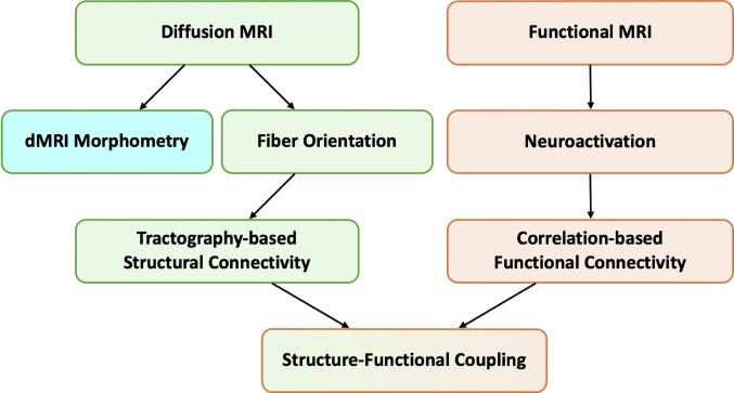
Traditional brain connectivity studies have focused mainly on structural connectivity, often relying on tractography with diffusion MRI (dMRI) to reconstruct white matter pathways. In parallel, studies of functional connectivity have examined correlations in brain activity using fMRI. However, emerging methodologies are advancing our understanding of brain networks. Here we explore advanced connectivity approaches beyond conventional tractography, focusing on dMRI morphometry and the integration of structural and functional connectivity analysis. dMRI morphometry enables quantitative assessment of white matter pathway volumes through statistical comparison with normative populations, while functional connectivity reveals network organization that is not restricted to direct anatomical connections. More recently, approaches that combine diffusion tensor imaging (DTI) with functional correlation tensor (FCT) analysis have been introduced, and these complementary methods provide new perspectives into brain structure-function relationships. Together, such approaches have important implications for neurodevelopmental and neurological disorders as well as brain plasticity. The integration of these methods with artificial intelligence techniques have the potential to support both basic neuroscience research and clinical applications.
Keywords: Functional MRI; Functional correlation tensor; Microstructure; Structure-function coupling; White matter.
© 2025. The Author(s).
Conflict of interest statement
Declarations. Conflict of interest: The authors declare no competing interests.
Figures

References
-
- Alexander DC, Dyrby TB, Nilsson M, Zhang H (2019) Imaging brain microstructure with diffusion MRI: practicality and applications. NMR Biomed 32:e3841. 10.1002/nbm.3841 - PubMed
-
- Beckmann CF, Smith SM (2004) Probabilistic independent component analysis for functional magnetic resonance imaging. IEEE Trans Méd Imaging 23:137–152. 10.1109/tmi.2003.822821 - PubMed
-
- Biswal B, Yetkin FZ, Haughton VM, Hyde JS (1995) Functional connectivity in the motor cortex of resting human brain using echo-planar mri. Magn Reson Med 34:537–541. 10.1002/mrm.1910340409 - PubMed
Publication types
MeSH terms
LinkOut - more resources
Full Text Sources
